 Movie
Movie Controller


+ Open data

- Basic information
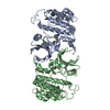
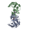
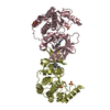
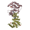
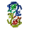
| Entry | Database: PDB / ID: 6gzm | ||||||
|---|---|---|---|---|---|---|---|
| Title | Crystal Structure of Human CKIdelta with A86 | ||||||
 Components Components | Casein kinase I isoform delta | ||||||
 Keywords Keywords | TRANSFERASE / CDK9 / kinase inhibitor / A86 | ||||||
| Function / homology |  Function and homology information Function and homology informationpositive regulation of non-canonical Wnt signaling pathway / protein localization to Golgi apparatus / COPII vesicle coat assembly / tau-protein kinase / microtubule nucleation / protein localization to cilium / The CRY:PER:kinase complex represses transactivation by the BMAL:CLOCK (ARNTL:CLOCK) complex / midbrain dopaminergic neuron differentiation / non-motile cilium assembly / protein localization to centrosome ...positive regulation of non-canonical Wnt signaling pathway / protein localization to Golgi apparatus / COPII vesicle coat assembly / tau-protein kinase / microtubule nucleation / protein localization to cilium / The CRY:PER:kinase complex represses transactivation by the BMAL:CLOCK (ARNTL:CLOCK) complex / midbrain dopaminergic neuron differentiation / non-motile cilium assembly / protein localization to centrosome / COPII-mediated vesicle transport / Phosphorylation and nuclear translocation of the CRY:PER:kinase complex / tau-protein kinase activity / Golgi organization / Major pathway of rRNA processing in the nucleolus and cytosol / spindle assembly / endoplasmic reticulum-Golgi intermediate compartment membrane / Loss of Nlp from mitotic centrosomes / Loss of proteins required for interphase microtubule organization from the centrosome / Recruitment of mitotic centrosome proteins and complexes / Recruitment of NuMA to mitotic centrosomes / Anchoring of the basal body to the plasma membrane / AURKA Activation by TPX2 / spindle microtubule / circadian regulation of gene expression / sperm end piece / regulation of circadian rhythm / spindle / Wnt signaling pathway / endocytosis / Regulation of PLK1 Activity at G2/M Transition / positive regulation of canonical Wnt signaling pathway / positive regulation of proteasomal ubiquitin-dependent protein catabolic process / actin cytoskeleton / sperm principal piece / protein phosphorylation / protein kinase activity / non-specific serine/threonine protein kinase / cilium / ciliary basal body / cadherin binding / protein serine kinase activity / protein serine/threonine kinase activity / centrosome / perinuclear region of cytoplasm / Golgi apparatus / signal transduction / nucleoplasm / ATP binding / nucleus / plasma membrane / cytoplasm / cytosol Similarity search - Function | ||||||
| Biological species |  Homo sapiens (human) Homo sapiens (human) | ||||||
| Method |  X-RAY DIFFRACTION / SYNCHROTRON / MOLECULAR REPLACEMENT / Resolution: 1.59 Å X-RAY DIFFRACTION / SYNCHROTRON / MOLECULAR REPLACEMENT / Resolution: 1.59 Å | ||||||
 Authors Authors | Ben-neriah, Y. / Venkatachalam, A. / Minzel, W. / Fink, A. / Snir-Alkalay, I. / Vacca, J. | ||||||
 Citation Citation | Journal: Cell / Year: 2018 Title: Small Molecules Co-targeting CKI alpha and the Transcriptional Kinases CDK7/9 Control AML in Preclinical Models. Authors: Minzel, W. / Venkatachalam, A. / Fink, A. / Hung, E. / Brachya, G. / Burstain, I. / Shaham, M. / Rivlin, A. / Omer, I. / Zinger, A. / Elias, S. / Winter, E. / Erdman, P.E. / Sullivan, R.W. / ...Authors: Minzel, W. / Venkatachalam, A. / Fink, A. / Hung, E. / Brachya, G. / Burstain, I. / Shaham, M. / Rivlin, A. / Omer, I. / Zinger, A. / Elias, S. / Winter, E. / Erdman, P.E. / Sullivan, R.W. / Fung, L. / Mercurio, F. / Li, D. / Vacca, J. / Kaushansky, N. / Shlush, L. / Oren, M. / Levine, R. / Pikarsky, E. / Snir-Alkalay, I. / Ben-Neriah, Y. | ||||||
| History |
|
- Structure visualization
| Structure viewer | Molecule: MolmilJmol/JSmol |
|---|
- Downloads & links
-Download
| PDBx/mmCIF format | 6gzm.cif.gz | 162.4 KB | Display | PDBx/mmCIF format |
|---|---|---|---|---|
| PDB format | pdb6gzm.ent.gz | 127.2 KB | Display | PDB format |
| PDBx/mmJSON format | 6gzm.json.gz | Tree view | PDBx/mmJSON format | |
| Others |  Other downloads Other downloads |
-Validation report
| Arichive directory | https://data.pdbj.org/pub/pdb/validation_reports/gz/6gzmftp://data.pdbj.org/pub/pdb/validation_reports/gz/6gzm | HTTPS FTP |
|---|
-Related structure data
| Related structure data |  6gzdC  6gzhC  3uytS S: Starting model for refinement C: citing same article ( |
|---|---|
| Similar structure data |


-Links
 PDBj
PDBj


- Assembly
| Deposited unit | 
| ||||||||
|---|---|---|---|---|---|---|---|---|---|
| 1 |
| ||||||||
| Unit cell |
|
-Components
| #1: Protein | Mass: 34310.613 Da / Num. of mol.: 2 Source method: isolated from a genetically manipulated source Source: (gene. exp.) Homo sapiens (human) / Gene: CSNK1D, HCKID / Production host:  References: UniProt: P48730, non-specific serine/threonine protein kinase, tau-protein kinase #2: Chemical | ChemComp-CIT / |   Mass: 192.124 Da / Num. of mol.: 1 / Source method: obtained synthetically / Formula: C6H8O7 Mass: 192.124 Da / Num. of mol.: 1 / Source method: obtained synthetically / Formula: C6H8O7#3: Chemical | ChemComp-GOL /   Mass: 92.094 Da / Num. of mol.: 4 / Source method: obtained synthetically / Formula: C3H8O3 Mass: 92.094 Da / Num. of mol.: 4 / Source method: obtained synthetically / Formula: C3H8O3#4: Chemical | 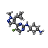  Mass: 345.438 Da / Num. of mol.: 2 / Source method: obtained synthetically / Formula: C18H26FN6 / Feature type: SUBJECT OF INVESTIGATION Mass: 345.438 Da / Num. of mol.: 2 / Source method: obtained synthetically / Formula: C18H26FN6 / Feature type: SUBJECT OF INVESTIGATION#5: Water | ChemComp-HOH / |  Mass: 18.015 Da / Num. of mol.: 832 / Source method: isolated from a natural source / Formula: H2O Mass: 18.015 Da / Num. of mol.: 832 / Source method: isolated from a natural source / Formula: H2O |
|---|
-Experimental details
-Experiment
| Experiment | Method: X-RAY DIFFRACTION / Number of used crystals: 1 |
|---|
- Sample preparation
| Crystal | Density Matthews: 2.29 Å3/Da / Density % sol: 46.32 % |
|---|---|
| Crystal grow | Temperature: 293.15 K / Method: vapor diffusion, hanging drop / pH: 7.5 Details: The construct used for crystallization was that of the wild type protein with the mutation R13N. Crystals of CK1d in complex with A-86 were obtained using hanging drop vapour diffusion set- ...Details: The construct used for crystallization was that of the wild type protein with the mutation R13N. Crystals of CK1d in complex with A-86 were obtained using hanging drop vapour diffusion set-ups. CK1d at a concentration of 13.6 mg/ml (50 mM HEPES, 266 mM NaCl, 1 mM EDTA, 1 mM DTT, 5 mM B-OG, pH 7.5) was pre-incubated with 2 mM (5.1-fold molar excess) of A-86 (150 mM in DMSO) for 1 h. 1 ul of the protein solution was then mixed with 1 ul of reservoir solution (0.1 M Na3-Citrate, pH 4.9, 18 %(w/v) PEG 3350) and equilibrated at 20 C over 0.4 ml of reservoir solution. Well diffracting crystals appeared over night |
-Data collection
| Diffraction | Mean temperature: 110.15 K |
|---|---|
| Diffraction source | Source: SYNCHROTRON / Site: ESRF  / Beamline: MASSIF-1 / Wavelength: 0.966 Å / Beamline: MASSIF-1 / Wavelength: 0.966 Å |
| Detector | Type: DECTRIS PILATUS3 2M / Detector: PIXEL / Date: Dec 19, 2016 |
| Radiation | Protocol: SINGLE WAVELENGTH / Monochromatic (M) / Laue (L): M / Scattering type: x-ray |
| Radiation wavelength | Wavelength: 0.966 Å / Relative weight: 1 |
| Reflection | Resolution: 1.59→29.19 Å / Num. obs: 80180 / % possible obs: 96.5 % / Redundancy: 2.4 % / Rrim(I) all: 0.09 / Net I/σ(I): 7.1 |
| Reflection shell | Resolution: 1.59→1.68 Å / Redundancy: 2.4 % / Num. unique obs: 11624 / Rrim(I) all: 0.57 / % possible all: 96.4 |
- Processing
| Software |
| ||||||||||||||||||||||||||||||||||||||||||||||||||||||||||||||||||||||||||||||||||||||||||||||||||||||||||||||||||||||||||||||||||||||||||||||||||||||||||||||||||||||||||||||||||||||
|---|---|---|---|---|---|---|---|---|---|---|---|---|---|---|---|---|---|---|---|---|---|---|---|---|---|---|---|---|---|---|---|---|---|---|---|---|---|---|---|---|---|---|---|---|---|---|---|---|---|---|---|---|---|---|---|---|---|---|---|---|---|---|---|---|---|---|---|---|---|---|---|---|---|---|---|---|---|---|---|---|---|---|---|---|---|---|---|---|---|---|---|---|---|---|---|---|---|---|---|---|---|---|---|---|---|---|---|---|---|---|---|---|---|---|---|---|---|---|---|---|---|---|---|---|---|---|---|---|---|---|---|---|---|---|---|---|---|---|---|---|---|---|---|---|---|---|---|---|---|---|---|---|---|---|---|---|---|---|---|---|---|---|---|---|---|---|---|---|---|---|---|---|---|---|---|---|---|---|---|---|---|---|---|
| Refinement | Method to determine structure: MOLECULAR REPLACEMENT Starting model: 3uyt Resolution: 1.59→29.19 Å / Cor.coef. Fo:Fc: 0.964 / Cor.coef. Fo:Fc free: 0.922 / SU B: 3.08 / SU ML: 0.104 / Cross valid method: THROUGHOUT / ESU R: 0.107 / ESU R Free: 0.117 / Stereochemistry target values: MAXIMUM LIKELIHOOD / Details: HYDROGENS HAVE BEEN ADDED IN THE RIDING POSITIONS
| ||||||||||||||||||||||||||||||||||||||||||||||||||||||||||||||||||||||||||||||||||||||||||||||||||||||||||||||||||||||||||||||||||||||||||||||||||||||||||||||||||||||||||||||||||||||
| Solvent computation | Ion probe radii: 0.8 Å / Shrinkage radii: 0.8 Å / VDW probe radii: 1.2 Å / Solvent model: BABINET MODEL WITH MASK | ||||||||||||||||||||||||||||||||||||||||||||||||||||||||||||||||||||||||||||||||||||||||||||||||||||||||||||||||||||||||||||||||||||||||||||||||||||||||||||||||||||||||||||||||||||||
| Displacement parameters | Biso mean: 28.076 Å2
| ||||||||||||||||||||||||||||||||||||||||||||||||||||||||||||||||||||||||||||||||||||||||||||||||||||||||||||||||||||||||||||||||||||||||||||||||||||||||||||||||||||||||||||||||||||||
| Refinement step | Cycle: 1 / Resolution: 1.59→29.19 Å
| ||||||||||||||||||||||||||||||||||||||||||||||||||||||||||||||||||||||||||||||||||||||||||||||||||||||||||||||||||||||||||||||||||||||||||||||||||||||||||||||||||||||||||||||||||||||
| Refine LS restraints |
|