+ データを開く
- 基本情報
基本情報
| 登録情報 | データベース: PDB / ID: 6gy7 | ||||||
|---|---|---|---|---|---|---|---|
| タイトル | Crystal structure of XaxB from Xenorhabdus nematophil | ||||||
 要素 要素 | XaxB | ||||||
 キーワード キーワード | TOXIN / bacterial toxin / pore forming-toxins | ||||||
| 機能・相同性 | : / XaxB 機能・相同性情報 機能・相同性情報 | ||||||
| 生物種 |  Xenorhabdus nematophila (バクテリア) Xenorhabdus nematophila (バクテリア) | ||||||
| 手法 |  X線回折 / X線回折 /  シンクロトロン / シンクロトロン /  単波長異常分散 / 解像度: 3.4 Å 単波長異常分散 / 解像度: 3.4 Å | ||||||
 データ登録者 データ登録者 | Schubert, E. / Raunser, S. / Vetter, I.R. / Prumbaum, D. / Penczek, P.A. | ||||||
| 資金援助 |  ドイツ, 1件 ドイツ, 1件
| ||||||
 引用 引用 |  ジャーナル: Elife / 年: 2018 ジャーナル: Elife / 年: 2018タイトル: Membrane insertion of α-xenorhabdolysin in near-atomic detail. 著者: Evelyn Schubert / Ingrid R Vetter / Daniel Prumbaum / Pawel A Penczek / Stefan Raunser /   要旨: α-Xenorhabdolysins (Xax) are α-pore-forming toxins (α-PFT) that form 1-1.3 MDa large pore complexes to perforate the host cell membrane. PFTs are used by a variety of bacterial pathogens to attack ...α-Xenorhabdolysins (Xax) are α-pore-forming toxins (α-PFT) that form 1-1.3 MDa large pore complexes to perforate the host cell membrane. PFTs are used by a variety of bacterial pathogens to attack host cells. Due to the lack of structural information, the molecular mechanism of action of Xax toxins is poorly understood. Here, we report the cryo-EM structure of the XaxAB pore complex from and the crystal structures of the soluble monomers of XaxA and XaxB. The structures reveal that XaxA and XaxB are built similarly and appear as heterodimers in the 12-15 subunits containing pore, classifying XaxAB as bi-component α-PFT. Major conformational changes in XaxB, including the swinging out of an amphipathic helix are responsible for membrane insertion. XaxA acts as an activator and stabilizer for XaxB that forms the actual transmembrane pore. Based on our results, we propose a novel structural model for the mechanism of Xax intoxication. | ||||||
| 履歴 |
|
- 構造の表示
構造の表示
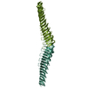
| 構造ビューア | 分子:  Molmil Molmil Jmol/JSmol Jmol/JSmol |
|---|
- ダウンロードとリンク
- ダウンロード
| PDBx/mmCIF形式 |  6gy7.cif.gz 6gy7.cif.gz | 495.5 KB | 表示 |  PDBx/mmCIF形式 PDBx/mmCIF形式 |
|---|---|---|---|---|
| PDB形式 |  pdb6gy7.ent.gz pdb6gy7.ent.gz | 418.6 KB | 表示 |  PDB形式 PDB形式 |
| PDBx/mmJSON形式 |  6gy7.json.gz 6gy7.json.gz | ツリー表示 |  PDBx/mmJSON形式 PDBx/mmJSON形式 | |
| その他 |  その他のダウンロード その他のダウンロード |
-検証レポート
| 文書・要旨 |  6gy7_validation.pdf.gz 6gy7_validation.pdf.gz | 457.2 KB | 表示 |  wwPDB検証レポート wwPDB検証レポート |
|---|---|---|---|---|
| 文書・詳細版 |  6gy7_full_validation.pdf.gz 6gy7_full_validation.pdf.gz | 470.4 KB | 表示 | |
| XML形式データ |  6gy7_validation.xml.gz 6gy7_validation.xml.gz | 42.9 KB | 表示 | |
| CIF形式データ |  6gy7_validation.cif.gz 6gy7_validation.cif.gz | 58.6 KB | 表示 | |
| アーカイブディレクトリ |  https://data.pdbj.org/pub/pdb/validation_reports/gy/6gy7 https://data.pdbj.org/pub/pdb/validation_reports/gy/6gy7 ftp://data.pdbj.org/pub/pdb/validation_reports/gy/6gy7 ftp://data.pdbj.org/pub/pdb/validation_reports/gy/6gy7 | HTTPS FTP |
-関連構造データ
- リンク
リンク
- 集合体
集合体
| 登録構造単位 | 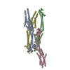
| ||||||||
|---|---|---|---|---|---|---|---|---|---|
| 1 | 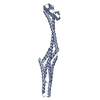
| ||||||||
| 2 | 
| ||||||||
| 3 | 
| ||||||||
| 4 | 
| ||||||||
| 単位格子 |
|
- 要素
要素
| #1: タンパク質 | 分子量: 41885.395 Da / 分子数: 4 / 由来タイプ: 組換発現 由来: (組換発現)  Xenorhabdus nematophila (strain ATCC 19061 / DSM 3370 / LMG 1036 / NCIB 9965 / AN6) (バクテリア) Xenorhabdus nematophila (strain ATCC 19061 / DSM 3370 / LMG 1036 / NCIB 9965 / AN6) (バクテリア)遺伝子: xaxB, XNC1_3767 / 発現宿主:  |
|---|
-実験情報
-実験
| 実験 | 手法:  X線回折 / 使用した結晶の数: 1 X線回折 / 使用した結晶の数: 1 |
|---|
- 試料調製
試料調製
| 結晶 | マシュー密度: 2.55 Å3/Da / 溶媒含有率: 51.85 % |
|---|---|
| 結晶化 | 温度: 293.15 K / 手法: 蒸気拡散法, シッティングドロップ法 / pH: 7.5 / 詳細: 0.2 M NaBr, 0.1 KCl, 20 % PEG 3350 |
-データ収集
| 回折 | 平均測定温度: 100 K |
|---|---|
| 放射光源 | 由来:  シンクロトロン / サイト: シンクロトロン / サイト:  SLS SLS  / ビームライン: X10SA / 波長: 0.97793 Å / ビームライン: X10SA / 波長: 0.97793 Å |
| 検出器 | タイプ: PSI PILATUS 6M / 検出器: PIXEL / 日付: 2018年2月24日 |
| 放射 | プロトコル: SINGLE WAVELENGTH / 単色(M)・ラウエ(L): M / 散乱光タイプ: x-ray |
| 放射波長 | 波長: 0.97793 Å / 相対比: 1 |
| 反射 | 解像度: 3.4→50 Å / Num. obs: 45577 / % possible obs: 100 % / 冗長度: 21.1 % / Net I/σ(I): 10.36 |
| 反射 シェル | 解像度: 3.4→3.49 Å |
- 解析
解析
| ソフトウェア |
| ||||||||||||||||||||||||||||||||||||||||||||||||||||||||||||||||||||||
|---|---|---|---|---|---|---|---|---|---|---|---|---|---|---|---|---|---|---|---|---|---|---|---|---|---|---|---|---|---|---|---|---|---|---|---|---|---|---|---|---|---|---|---|---|---|---|---|---|---|---|---|---|---|---|---|---|---|---|---|---|---|---|---|---|---|---|---|---|---|---|---|
| 精密化 | 構造決定の手法:  単波長異常分散 / 解像度: 3.4→48.537 Å / SU ML: 0.55 / 交差検証法: FREE R-VALUE / σ(F): 1.34 / 位相誤差: 35.77 / 立体化学のターゲット値: ML 単波長異常分散 / 解像度: 3.4→48.537 Å / SU ML: 0.55 / 交差検証法: FREE R-VALUE / σ(F): 1.34 / 位相誤差: 35.77 / 立体化学のターゲット値: ML
| ||||||||||||||||||||||||||||||||||||||||||||||||||||||||||||||||||||||
| 溶媒の処理 | 減衰半径: 0.9 Å / VDWプローブ半径: 1.11 Å / 溶媒モデル: FLAT BULK SOLVENT MODEL | ||||||||||||||||||||||||||||||||||||||||||||||||||||||||||||||||||||||
| 精密化ステップ | サイクル: LAST / 解像度: 3.4→48.537 Å
| ||||||||||||||||||||||||||||||||||||||||||||||||||||||||||||||||||||||
| 拘束条件 |
| ||||||||||||||||||||||||||||||||||||||||||||||||||||||||||||||||||||||
| LS精密化 シェル |
|
 ムービー
ムービー コントローラー


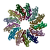


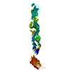


 PDBj
PDBj