 Movie
Movie Controller


+ Open data

- Basic information
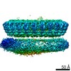
| Entry | Database: PDB / ID: 6gy6 | ||||||
|---|---|---|---|---|---|---|---|
| Title | XaxAB pore complex from Xenorhabdus nematophila | ||||||
 Components Components |
| ||||||
 Keywords Keywords | TOXIN / bacterial toxin / pore forming-toxins | ||||||
| Function / homology | : / XaxA / XaxB Function and homology information Function and homology information | ||||||
| Biological species |  Xenorhabdus nematophila ATCC 19061 (bacteria) Xenorhabdus nematophila ATCC 19061 (bacteria) | ||||||
| Method | ELECTRON MICROSCOPY / single particle reconstruction / cryo EM / Resolution: 4 Å | ||||||
 Authors Authors | Schubert, E. / Raunser, S. | ||||||
| Funding support |  Germany, 1items Germany, 1items
| ||||||
 Citation Citation | Journal: Elife / Year: 2018 Title: Membrane insertion of α-xenorhabdolysin in near-atomic detail. Authors: Evelyn Schubert / Ingrid R Vetter / Daniel Prumbaum / Pawel A Penczek / Stefan Raunser /  Abstract: α-Xenorhabdolysins (Xax) are α-pore-forming toxins (α-PFT) that form 1-1.3 MDa large pore complexes to perforate the host cell membrane. PFTs are used by a variety of bacterial pathogens to attack ...α-Xenorhabdolysins (Xax) are α-pore-forming toxins (α-PFT) that form 1-1.3 MDa large pore complexes to perforate the host cell membrane. PFTs are used by a variety of bacterial pathogens to attack host cells. Due to the lack of structural information, the molecular mechanism of action of Xax toxins is poorly understood. Here, we report the cryo-EM structure of the XaxAB pore complex from and the crystal structures of the soluble monomers of XaxA and XaxB. The structures reveal that XaxA and XaxB are built similarly and appear as heterodimers in the 12-15 subunits containing pore, classifying XaxAB as bi-component α-PFT. Major conformational changes in XaxB, including the swinging out of an amphipathic helix are responsible for membrane insertion. XaxA acts as an activator and stabilizer for XaxB that forms the actual transmembrane pore. Based on our results, we propose a novel structural model for the mechanism of Xax intoxication. | ||||||
| History |
|
- Structure visualization
| Movie |
Movie viewer |
|---|---|
| Structure viewer | Molecule: MolmilJmol/JSmol |
- Downloads & links
-Download
| PDBx/mmCIF format | 6gy6.cif.gz | 1.5 MB | Display | PDBx/mmCIF format |
|---|---|---|---|---|
| PDB format | pdb6gy6.ent.gz | 1.3 MB | Display | PDB format |
| PDBx/mmJSON format | 6gy6.json.gz | Tree view | PDBx/mmJSON format | |
| Others |  Other downloads Other downloads |
-Validation report
| Arichive directory | https://data.pdbj.org/pub/pdb/validation_reports/gy/6gy6ftp://data.pdbj.org/pub/pdb/validation_reports/gy/6gy6 | HTTPS FTP |
|---|
-Related structure data
| Related structure data | 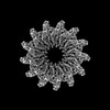 0088MC  6gy7C  6gy8C M: map data used to model this data C: citing same article ( |
|---|---|
| Similar structure data |

-Links
 PDBj
PDBj- Assembly
| Deposited unit | 
|
|---|---|
| 1 |
|
-Components
| #1: Protein | Mass: 47465.859 Da / Num. of mol.: 13 Source method: isolated from a genetically manipulated source Source: (gene. exp.) Xenorhabdus nematophila ATCC 19061 (bacteria)Gene: xaxA, XNC1_3766 / Production host: #2: Protein | Mass: 38518.688 Da / Num. of mol.: 13 Source method: isolated from a genetically manipulated source Source: (gene. exp.) Xenorhabdus nematophila ATCC 19061 (bacteria)Gene: xaxB, XNC1_3767 / Production host: Has protein modification | N | |
|---|
-Experimental details
-Experiment
| Experiment | Method: ELECTRON MICROSCOPY |
|---|---|
| EM experiment | Aggregation state: PARTICLE / 3D reconstruction method: single particle reconstruction |
- Sample preparation
| Component |
| ||||||||||||||||||||||||||||
|---|---|---|---|---|---|---|---|---|---|---|---|---|---|---|---|---|---|---|---|---|---|---|---|---|---|---|---|---|---|
| Molecular weight |
| ||||||||||||||||||||||||||||
| Source (natural) |
| ||||||||||||||||||||||||||||
| Source (recombinant) |
| ||||||||||||||||||||||||||||
| Buffer solution | pH: 7.5 | ||||||||||||||||||||||||||||
| Specimen | Conc.: 1 mg/ml / Embedding applied: NO / Shadowing applied: NO / Staining applied: NO / Vitrification applied: YES | ||||||||||||||||||||||||||||
| Specimen support | Grid type: C-flat-2/1 | ||||||||||||||||||||||||||||
| Vitrification | Instrument: GATAN CRYOPLUNGE 3 / Cryogen name: ETHANE / Humidity: 90 % / Chamber temperature: 298.15 K |
- Electron microscopy imaging
| Experimental equipment |  Model: Titan Krios / Image courtesy: FEI Company |
|---|---|
| Microscopy | Model: FEI TITAN KRIOS |
| Electron gun | Electron source:  FIELD EMISSION GUN / Accelerating voltage: 300 kV / Illumination mode: SPOT SCAN FIELD EMISSION GUN / Accelerating voltage: 300 kV / Illumination mode: SPOT SCAN |
| Electron lens | Mode: BRIGHT FIELD |
| Image recording | Electron dose: 44 e/Å2 / Detector mode: COUNTING / Film or detector model: FEI FALCON III (4k x 4k) |
- Processing
| EM software |
| ||||||||||||||||||||
|---|---|---|---|---|---|---|---|---|---|---|---|---|---|---|---|---|---|---|---|---|---|
| CTF correction | Type: PHASE FLIPPING AND AMPLITUDE CORRECTION | ||||||||||||||||||||
| Symmetry | Point symmetry: C13 (13 fold cyclic) | ||||||||||||||||||||
| 3D reconstruction | Resolution: 4 Å / Resolution method: FSC 0.143 CUT-OFF / Num. of particles: 43305 / Symmetry type: POINT |
