 Movie
Movie Controller

[English] 日本語
 Yorodumi
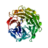
- PDB-6gxs: Crystal structure of CV39L lectin from Chromobacterium violaceum ... -
+ Open data

- Basic information
| Entry | Database: PDB / ID: 6gxs | |||||||||
|---|---|---|---|---|---|---|---|---|---|---|
| Title | Crystal structure of CV39L lectin from Chromobacterium violaceum at 1.8 A resolution | |||||||||
 Components Components | CV39L lectin | |||||||||
 Keywords Keywords | SUGAR BINDING PROTEIN / Lectin | |||||||||
| Function / homology | DI(HYDROXYETHYL)ETHER / Uncharacterized protein Function and homology information Function and homology information | |||||||||
| Biological species |  Chromobacterium violaceum (bacteria) Chromobacterium violaceum (bacteria) | |||||||||
| Method |  X-RAY DIFFRACTION / SYNCHROTRON / SAD / Resolution: 1.8 Å X-RAY DIFFRACTION / SYNCHROTRON / SAD / Resolution: 1.8 Å | |||||||||
 Authors Authors | Sykorova, P. / Novotna, J. / Demo, G. / Pompidor, G. / Dubska, E. / Komarek, J. / Fujdiarova, E. / Haronikova, L. / Varrot, A. / Imberty, A. ...Sykorova, P. / Novotna, J. / Demo, G. / Pompidor, G. / Dubska, E. / Komarek, J. / Fujdiarova, E. / Haronikova, L. / Varrot, A. / Imberty, A. / Shilova, N. / Bovin, N. / Pokorna, M. / Wimmerova, M. | |||||||||
| Funding support |  Czech Republic, 2items Czech Republic, 2items
| |||||||||
 Citation Citation | Journal: Int.J.Biol.Macromol. / Year: 2020 Title: Characterization of novel lectins from Burkholderia pseudomallei and Chromobacterium violaceum with seven-bladed beta-propeller fold. Authors: Sykorova, P. / Novotna, J. / Demo, G. / Pompidor, G. / Dubska, E. / Komarek, J. / Fujdiarova, E. / Houser, J. / Haronikova, L. / Varrot, A. / Shilova, N. / Imberty, A. / Bovin, N. / Pokorna, M. / Wimmerova, M. | |||||||||
| History |
|
- Structure visualization
| Structure viewer | Molecule: MolmilJmol/JSmol |
|---|
- Downloads & links
-Download
| PDBx/mmCIF format | 6gxs.cif.gz | 303.4 KB | Display | PDBx/mmCIF format |
|---|---|---|---|---|
| PDB format | pdb6gxs.ent.gz | 242.3 KB | Display | PDB format |
| PDBx/mmJSON format | 6gxs.json.gz | Tree view | PDBx/mmJSON format | |
| Others |  Other downloads Other downloads |
-Validation report
| Arichive directory | https://data.pdbj.org/pub/pdb/validation_reports/gx/6gxsftp://data.pdbj.org/pub/pdb/validation_reports/gx/6gxs | HTTPS FTP |
|---|
-Related structure data


-Links
 PDBj
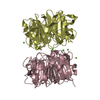
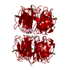
PDBj- Assembly
| Deposited unit | 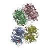
| ||||||||
|---|---|---|---|---|---|---|---|---|---|
| 1 | 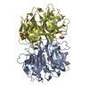
| ||||||||
| 2 | 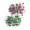
| ||||||||
| Unit cell |
|
-Components
-Protein , 1 types, 4 molecules ABCD
| #1: Protein | Mass: 39811.203 Da / Num. of mol.: 4 Source method: isolated from a genetically manipulated source Source: (gene. exp.) Chromobacterium violaceum (strain ATCC 12472 / DSM 30191 / JCM 1249 / NBRC 12614 / NCIMB 9131 / NCTC 9757) (bacteria)Gene: CV_1052 / Production host: |
|---|
-Non-polymers , 5 types, 1292 molecules 


| #2: Chemical | ChemComp-SO4 /  Mass: 96.063 Da / Num. of mol.: 11 / Source method: obtained synthetically / Formula: SO4 Mass: 96.063 Da / Num. of mol.: 11 / Source method: obtained synthetically / Formula: SO4#3: Chemical |  Mass: 62.068 Da / Num. of mol.: 2 / Source method: obtained synthetically / Formula: C2H6O2 Mass: 62.068 Da / Num. of mol.: 2 / Source method: obtained synthetically / Formula: C2H6O2#4: Chemical |  Mass: 106.120 Da / Num. of mol.: 3 / Source method: obtained synthetically / Formula: C4H10O3 Mass: 106.120 Da / Num. of mol.: 3 / Source method: obtained synthetically / Formula: C4H10O3#5: Chemical | ChemComp-1PE / |  Mass: 238.278 Da / Num. of mol.: 1 / Source method: obtained synthetically / Formula: C10H22O6 / Comment: precipitant*YM Mass: 238.278 Da / Num. of mol.: 1 / Source method: obtained synthetically / Formula: C10H22O6 / Comment: precipitant*YM#6: Water | ChemComp-HOH / | Mass: 18.015 Da / Num. of mol.: 1275 / Source method: isolated from a natural source / Formula: H2O |
|---|
-Experimental details
-Experiment
| Experiment | Method: X-RAY DIFFRACTION / Number of used crystals: 1 |
|---|
- Sample preparation
| Crystal | Density Matthews: 2.18 Å3/Da / Density meas: 1471639.375 Mg/m3 / Density % sol: 43.6 % |
|---|---|
| Crystal grow | Temperature: 290 K / Method: vapor diffusion, hanging drop / pH: 4.6 Details: 0.1 M sodium acetate, 25% PEG 4000, 0.2M ammonium sulphate, pH 4.6 |
-Data collection
| Diffraction | Mean temperature: 100 K |
|---|---|
| Diffraction source | Source: SYNCHROTRON / Site: PETRA III, EMBL c/o DESY  / Beamline: P13 (MX1) / Wavelength: 0.967 Å / Beamline: P13 (MX1) / Wavelength: 0.967 Å |
| Detector | Type: DECTRIS PILATUS3 S 6M / Detector: PIXEL / Date: Oct 31, 2013 |
| Radiation | Protocol: SINGLE WAVELENGTH / Monochromatic (M) / Laue (L): M / Scattering type: x-ray |
| Radiation wavelength | Wavelength: 0.967 Å / Relative weight: 1 |
| Reflection | Resolution: 1.8→80.93 Å / Num. obs: 137285 / % possible obs: 100 % / Redundancy: 7.5 % / CC1/2: 0.999 / Rmerge(I) obs: 0.102 / Net I/σ(I): 14.7 |
| Reflection shell | Resolution: 1.8→1.9 Å / Redundancy: 7.5 % / Rmerge(I) obs: 0.363 / Mean I/σ(I) obs: 4.9 / Num. unique obs: 19822 / CC1/2: 0.953 / % possible all: 100 |
- Processing
| Software |
| ||||||||||||||||||||||||||||||||||||||||||||||||||||||||||||||||||||||||||||||||||||||||||||||||||||||||||||||||||||||||||||||||||||||||||||||||||||||||||||||||||||||||||||||||||||||
|---|---|---|---|---|---|---|---|---|---|---|---|---|---|---|---|---|---|---|---|---|---|---|---|---|---|---|---|---|---|---|---|---|---|---|---|---|---|---|---|---|---|---|---|---|---|---|---|---|---|---|---|---|---|---|---|---|---|---|---|---|---|---|---|---|---|---|---|---|---|---|---|---|---|---|---|---|---|---|---|---|---|---|---|---|---|---|---|---|---|---|---|---|---|---|---|---|---|---|---|---|---|---|---|---|---|---|---|---|---|---|---|---|---|---|---|---|---|---|---|---|---|---|---|---|---|---|---|---|---|---|---|---|---|---|---|---|---|---|---|---|---|---|---|---|---|---|---|---|---|---|---|---|---|---|---|---|---|---|---|---|---|---|---|---|---|---|---|---|---|---|---|---|---|---|---|---|---|---|---|---|---|---|---|
| Refinement | Method to determine structure: SAD / Resolution: 1.8→73.04 Å / Cor.coef. Fo:Fc: 0.957 / Cor.coef. Fo:Fc free: 0.942 / SU B: 1.661 / SU ML: 0.054 / Cross valid method: THROUGHOUT / ESU R: 0.023 / ESU R Free: 0.022 / Details: HYDROGENS HAVE BEEN ADDED IN THE RIDING POSITIONS
| ||||||||||||||||||||||||||||||||||||||||||||||||||||||||||||||||||||||||||||||||||||||||||||||||||||||||||||||||||||||||||||||||||||||||||||||||||||||||||||||||||||||||||||||||||||||
| Solvent computation | Ion probe radii: 0.8 Å / Shrinkage radii: 0.8 Å / VDW probe radii: 1.2 Å | ||||||||||||||||||||||||||||||||||||||||||||||||||||||||||||||||||||||||||||||||||||||||||||||||||||||||||||||||||||||||||||||||||||||||||||||||||||||||||||||||||||||||||||||||||||||
| Displacement parameters | Biso mean: 12.009 Å2
| ||||||||||||||||||||||||||||||||||||||||||||||||||||||||||||||||||||||||||||||||||||||||||||||||||||||||||||||||||||||||||||||||||||||||||||||||||||||||||||||||||||||||||||||||||||||
| Refinement step | Cycle: 1 / Resolution: 1.8→73.04 Å
| ||||||||||||||||||||||||||||||||||||||||||||||||||||||||||||||||||||||||||||||||||||||||||||||||||||||||||||||||||||||||||||||||||||||||||||||||||||||||||||||||||||||||||||||||||||||
| Refine LS restraints |
|