 Movie
Movie Controller


+ Open data

- Basic information
| Entry | Database: PDB / ID: 6rgj | ||||||
|---|---|---|---|---|---|---|---|
| Title | Photorhabdus asymbiotica lectin PHL in complex with D-glucose | ||||||
 Components Components | Lectin PHL | ||||||
 Keywords Keywords | SUGAR BINDING PROTEIN / lectin / Photorhabdus / glucose / beta-propeller | ||||||
| Function / homology | : / PLL-like beta propeller / Repeat of unknown function (DUF346) / metal ion binding / beta-D-glucopyranose / PLL-like beta propeller domain-containing protein Function and homology information Function and homology information | ||||||
| Biological species |  Photorhabdus asymbiotica (bacteria) Photorhabdus asymbiotica (bacteria) | ||||||
| Method |  X-RAY DIFFRACTION / SYNCHROTRON / MOLECULAR REPLACEMENT / Resolution: 1.8 Å X-RAY DIFFRACTION / SYNCHROTRON / MOLECULAR REPLACEMENT / Resolution: 1.8 Å | ||||||
 Authors Authors | Houser, J. / Fujdiarova, E. / Jancarikova, G. / Wimmerova, M. | ||||||
 Citation Citation | Journal: Febs J. / Year: 2021 Title: Heptabladed beta-propeller lectins PLL2 and PHL from Photorhabdus spp. recognize O-methylated sugars and influence the host immune system. Authors: Fujdiarova, E. / Houser, J. / Dobes, P. / Paulikova, G. / Kondakov, N. / Kononov, L. / Hyrsl, P. / Wimmerova, M. | ||||||
| History |
|
- Structure visualization
| Structure viewer | Molecule: MolmilJmol/JSmol |
|---|
- Downloads & links
-Download
| PDBx/mmCIF format | 6rgj.cif.gz | 93.1 KB | Display | PDBx/mmCIF format |
|---|---|---|---|---|
| PDB format | pdb6rgj.ent.gz | 67.7 KB | Display | PDB format |
| PDBx/mmJSON format | 6rgj.json.gz | Tree view | PDBx/mmJSON format | |
| Others |  Other downloads Other downloads |
-Validation report
| Arichive directory | https://data.pdbj.org/pub/pdb/validation_reports/rg/6rgjftp://data.pdbj.org/pub/pdb/validation_reports/rg/6rgj | HTTPS FTP |
|---|
-Related structure data
| Related structure data |  6rfzC  6rg1C  6rg2C 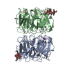 6rggC  6rgrC  6rguC  6rgwC  5mxeS S: Starting model for refinement C: citing same article ( |
|---|---|
| Similar structure data |


-Links
 PDBj
PDBj


- Assembly
| Deposited unit | 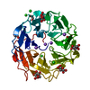
| |||||||||
|---|---|---|---|---|---|---|---|---|---|---|
| 1 | 
| |||||||||
| Unit cell |
| |||||||||
| Components on special symmetry positions |
|
-Components
-Protein / Sugars , 2 types, 5 molecules A
| #1: Protein | Mass: 40213.098 Da / Num. of mol.: 1 Source method: isolated from a genetically manipulated source Source: (gene. exp.) Photorhabdus asymbiotica (bacteria) / Gene: PAU_00698 / Plasmid: pET25-b / Production host: |
|---|---|
| #3: Sugar | ChemComp-BGC /  Type: D-saccharide, beta linking / Mass: 180.156 Da / Num. of mol.: 4 Type: D-saccharide, beta linking / Mass: 180.156 Da / Num. of mol.: 4Source method: isolated from a genetically manipulated source Formula: C6H12O6 |
-Non-polymers , 4 types, 369 molecules 


| #2: Chemical |  Mass: 62.068 Da / Num. of mol.: 3 / Source method: obtained synthetically / Formula: C2H6O2 Mass: 62.068 Da / Num. of mol.: 3 / Source method: obtained synthetically / Formula: C2H6O2#4: Chemical | ChemComp-NA /  Mass: 22.990 Da / Num. of mol.: 4 / Source method: obtained synthetically / Formula: Na Mass: 22.990 Da / Num. of mol.: 4 / Source method: obtained synthetically / Formula: Na#5: Chemical | ChemComp-CL /  Mass: 35.453 Da / Num. of mol.: 7 / Source method: obtained synthetically / Formula: Cl Mass: 35.453 Da / Num. of mol.: 7 / Source method: obtained synthetically / Formula: Cl#6: Water | ChemComp-HOH / | Mass: 18.015 Da / Num. of mol.: 355 / Source method: isolated from a natural source / Formula: H2O |
|---|
-Details
| Has protein modification | Y |
|---|
-Experimental details
-Experiment
| Experiment | Method: X-RAY DIFFRACTION / Number of used crystals: 1 |
|---|
- Sample preparation
| Crystal | Density Matthews: 2.84 Å3/Da / Density % sol: 56.69 % |
|---|---|
| Crystal grow | Temperature: 293 K / Method: vapor diffusion, hanging drop / pH: 7.5 / Details: 3.9 M NaCl, 100 mM Hepes, pH 7.5 |
-Data collection
| Diffraction | Mean temperature: 100 K / Serial crystal experiment: N |
|---|---|
| Diffraction source | Source: SYNCHROTRON / Site: BESSY  / Beamline: 14.3 / Wavelength: 0.895 Å / Beamline: 14.3 / Wavelength: 0.895 Å |
| Detector | Type: MARMOSAIC 300 mm CCD / Detector: CCD / Date: Aug 30, 2016 |
| Radiation | Protocol: SINGLE WAVELENGTH / Monochromatic (M) / Laue (L): M / Scattering type: x-ray |
| Radiation wavelength | Wavelength: 0.895 Å / Relative weight: 1 |
| Reflection | Resolution: 1.8→44.03 Å / Num. obs: 40112 / % possible obs: 99.7 % / Redundancy: 12.3 % / Biso Wilson estimate: 10.799 Å2 / CC1/2: 0.998 / Rmerge(I) obs: 0.144 / Net I/σ(I): 18.5 |
| Reflection shell | Resolution: 1.8→1.9 Å / Redundancy: 12.1 % / Rmerge(I) obs: 0.795 / Mean I/σ(I) obs: 4.5 / Num. unique obs: 5670 / CC1/2: 0.895 / % possible all: 98.2 |
- Processing
| Software |
| ||||||||||||||||||||||||||||||||||||||||||||||||||||||||||||||||||||||||||||||||||||||||||||||||||||||||||||||||||||||||||||||||||||||||||||||||||||||||||||||||||||||||||||||||||||||
|---|---|---|---|---|---|---|---|---|---|---|---|---|---|---|---|---|---|---|---|---|---|---|---|---|---|---|---|---|---|---|---|---|---|---|---|---|---|---|---|---|---|---|---|---|---|---|---|---|---|---|---|---|---|---|---|---|---|---|---|---|---|---|---|---|---|---|---|---|---|---|---|---|---|---|---|---|---|---|---|---|---|---|---|---|---|---|---|---|---|---|---|---|---|---|---|---|---|---|---|---|---|---|---|---|---|---|---|---|---|---|---|---|---|---|---|---|---|---|---|---|---|---|---|---|---|---|---|---|---|---|---|---|---|---|---|---|---|---|---|---|---|---|---|---|---|---|---|---|---|---|---|---|---|---|---|---|---|---|---|---|---|---|---|---|---|---|---|---|---|---|---|---|---|---|---|---|---|---|---|---|---|---|---|
| Refinement | Method to determine structure: MOLECULAR REPLACEMENT Starting model: 5MXE Resolution: 1.8→44.03 Å / Cor.coef. Fo:Fc: 0.958 / Cor.coef. Fo:Fc free: 0.948 / SU B: 2.38 / SU ML: 0.072 / Cross valid method: THROUGHOUT / ESU R: 0.108 / ESU R Free: 0.103 / Details: HYDROGENS HAVE BEEN ADDED IN THE RIDING POSITIONS
| ||||||||||||||||||||||||||||||||||||||||||||||||||||||||||||||||||||||||||||||||||||||||||||||||||||||||||||||||||||||||||||||||||||||||||||||||||||||||||||||||||||||||||||||||||||||
| Solvent computation | Ion probe radii: 0.8 Å / Shrinkage radii: 0.8 Å / VDW probe radii: 1.2 Å | ||||||||||||||||||||||||||||||||||||||||||||||||||||||||||||||||||||||||||||||||||||||||||||||||||||||||||||||||||||||||||||||||||||||||||||||||||||||||||||||||||||||||||||||||||||||
| Displacement parameters | Biso mean: 15.492 Å2
| ||||||||||||||||||||||||||||||||||||||||||||||||||||||||||||||||||||||||||||||||||||||||||||||||||||||||||||||||||||||||||||||||||||||||||||||||||||||||||||||||||||||||||||||||||||||
| Refinement step | Cycle: LAST / Resolution: 1.8→44.03 Å
| ||||||||||||||||||||||||||||||||||||||||||||||||||||||||||||||||||||||||||||||||||||||||||||||||||||||||||||||||||||||||||||||||||||||||||||||||||||||||||||||||||||||||||||||||||||||
| Refine LS restraints |
|