 Movie
Movie Controller

[English] 日本語
 Yorodumi
- PDB-4nc4: Crystal structure of photoreceptor AtUVR8 mutant W285F and light-... -
+ Open data

- Basic information
| Entry | Database: PDB / ID: 4nc4 | ||||||
|---|---|---|---|---|---|---|---|
| Title | Crystal structure of photoreceptor AtUVR8 mutant W285F and light-induced structural changes at 120K | ||||||
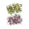
 Components Components | Ultraviolet-B receptor UVR8 | ||||||
 Keywords Keywords | SIGNALING PROTEIN / 7-blade beta-propeller | ||||||
| Function / homology |  Function and homology information Function and homology informationentrainment of circadian clock / response to UV-B / plastid / photoreceptor activity / response to UV / guanyl-nucleotide exchange factor activity / chromatin binding / chromatin / protein homodimerization activity / identical protein binding ...entrainment of circadian clock / response to UV-B / plastid / photoreceptor activity / response to UV / guanyl-nucleotide exchange factor activity / chromatin binding / chromatin / protein homodimerization activity / identical protein binding / nucleus / cytosol Similarity search - Function | ||||||
| Biological species |  | ||||||
| Method |  X-RAY DIFFRACTION / SYNCHROTRON / DIFFERENCE FOURIER / Resolution: 1.75 Å X-RAY DIFFRACTION / SYNCHROTRON / DIFFERENCE FOURIER / Resolution: 1.75 Å | ||||||
 Authors Authors | Yang, X. / Zeng, X. / Zhao, K.-H. / Ren, Z. | ||||||
 Citation Citation | Journal: Nat Plants / Year: 2015 Title: Dynamic Crystallography Reveals Early Signalling Events in Ultraviolet Photoreceptor UVR8. Authors: Zeng, X. / Ren, Z. / Wu, Q. / Fan, J. / Peng, P.P. / Tang, K. / Zhang, R. / Zhao, K.H. / Yang, X. | ||||||
| History |
|
- Structure visualization
| Structure viewer | Molecule: MolmilJmol/JSmol |
|---|
- Downloads & links
-Download
| PDBx/mmCIF format | 4nc4.cif.gz | 602.8 KB | Display | PDBx/mmCIF format |
|---|---|---|---|---|
| PDB format | pdb4nc4.ent.gz | 494.9 KB | Display | PDB format |
| PDBx/mmJSON format | 4nc4.json.gz | Tree view | PDBx/mmJSON format | |
| Others |  Other downloads Other downloads |
-Validation report
| Arichive directory | https://data.pdbj.org/pub/pdb/validation_reports/nc/4nc4ftp://data.pdbj.org/pub/pdb/validation_reports/nc/4nc4 | HTTPS FTP |
|---|
-Related structure data
| Related structure data |  4naaSC  4nbmC S: Starting model for refinement C: citing same article ( |
|---|---|
| Similar structure data |

-Links
 PDBj
PDBj- Assembly
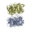
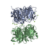
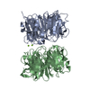
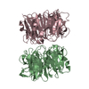
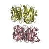
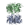
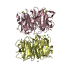
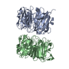
| Deposited unit | 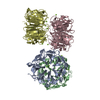
| |||||||||
|---|---|---|---|---|---|---|---|---|---|---|
| 1 | 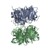
| |||||||||
| 2 | 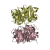
| |||||||||
| 3 | 
| |||||||||
| 4 | 
| |||||||||
| 5 | 
| |||||||||
| 6 | 
| |||||||||
| Unit cell |
| |||||||||
| Components on special symmetry positions |
|
-Components
| #1: Protein | Mass: 40836.398 Da / Num. of mol.: 4 / Fragment: UNP residues 13-382 / Mutation: W285F Source method: isolated from a genetically manipulated source Source: (gene. exp.)  #2: Chemical | ChemComp-MG /   Mass: 24.305 Da / Num. of mol.: 18 / Source method: obtained synthetically / Formula: Mg Mass: 24.305 Da / Num. of mol.: 18 / Source method: obtained synthetically / Formula: Mg#3: Water | ChemComp-HOH / |  Mass: 18.015 Da / Num. of mol.: 1643 / Source method: isolated from a natural source / Formula: H2O Mass: 18.015 Da / Num. of mol.: 1643 / Source method: isolated from a natural source / Formula: H2O |
|---|
-Experimental details
-Experiment
| Experiment | Method: X-RAY DIFFRACTION / Number of used crystals: 1 |
|---|
- Sample preparation
| Crystal | Density Matthews: 2.55 Å3/Da / Density % sol: 51.82 % |
|---|---|
| Crystal grow | Temperature: 290 K / pH: 9.2 Details: protein concentration 8mg/ml, mother liquor 0.1M MgCl2 15-18% PEG8000 and 0.1M Tris HCl pH 9.2, VAPOR DIFFUSION, HANGING DROP, temperature 290K |
-Data collection
| Diffraction | Mean temperature: 100 K |
|---|---|
| Diffraction source | Source: SYNCHROTRON / Site: APS  / Beamline: 21-ID-G / Wavelength: 0.97856 / Beamline: 21-ID-G / Wavelength: 0.97856 |
| Detector | Type: RAYONIX MX-300 / Detector: CCD / Date: Jul 5, 2013 |
| Radiation | Monochromator: DIAMOND LAUE MONOCHROMATOR / Protocol: SINGLE WAVELENGTH / Monochromatic (M) / Laue (L): M / Scattering type: x-ray |
| Radiation wavelength | Wavelength: 0.97856 Å / Relative weight: 1 |
| Reflection | Resolution: 1.75→50 Å / Num. obs: 164911 / % possible obs: 99.8 % / Observed criterion σ(I): 2 / Redundancy: 3.6 % / Rmerge(I) obs: 0.047 |
- Processing
| Software |
| |||||||||||||||||||||||||||||||||||||||||||||||||||||||||||||||||||||||||||||||||||||||||||||||||||||||||||||||||||||||||||||||||||||||||||||||||||||||||||||||||||||||||||||||||||||||||||||||||||||||||||||||||||||||||
|---|---|---|---|---|---|---|---|---|---|---|---|---|---|---|---|---|---|---|---|---|---|---|---|---|---|---|---|---|---|---|---|---|---|---|---|---|---|---|---|---|---|---|---|---|---|---|---|---|---|---|---|---|---|---|---|---|---|---|---|---|---|---|---|---|---|---|---|---|---|---|---|---|---|---|---|---|---|---|---|---|---|---|---|---|---|---|---|---|---|---|---|---|---|---|---|---|---|---|---|---|---|---|---|---|---|---|---|---|---|---|---|---|---|---|---|---|---|---|---|---|---|---|---|---|---|---|---|---|---|---|---|---|---|---|---|---|---|---|---|---|---|---|---|---|---|---|---|---|---|---|---|---|---|---|---|---|---|---|---|---|---|---|---|---|---|---|---|---|---|---|---|---|---|---|---|---|---|---|---|---|---|---|---|---|---|---|---|---|---|---|---|---|---|---|---|---|---|---|---|---|---|---|---|---|---|---|---|---|---|---|---|---|---|---|---|---|---|---|
| Refinement | Method to determine structure: DIFFERENCE FOURIER Starting model: 4NAA Resolution: 1.75→28.49 Å / SU ML: 0.16 / σ(F): 1.34 / Phase error: 25.21 / Stereochemistry target values: ML Details: THE AUTHOR STATES THAT ENTRIES 4NAA, 4NBM, AND 4NC4 WERE DERIVED FROM DYNAMIC CRYSTALLOGRAPHIC DATA SETS. SPECIFICALLY, EACH ENTRY PROVIDES THREE ASPECTS OF EXPERIMENTAL DATA THAT ARE NEEDED ...Details: THE AUTHOR STATES THAT ENTRIES 4NAA, 4NBM, AND 4NC4 WERE DERIVED FROM DYNAMIC CRYSTALLOGRAPHIC DATA SETS. SPECIFICALLY, EACH ENTRY PROVIDES THREE ASPECTS OF EXPERIMENTAL DATA THAT ARE NEEDED TO REPRODUCE AND VALIDATE OUR SCIENTIFIC FINDINGS. THEY INCLUDE: A) COORDINATES OF THE REFERENCE "DARK" STRUCTURE AS WE HAD DEPOSITED IN PDB; B) STRUCTURE FACTOR AMPLITUDES ACQUIRED IN THE "DARK" STATE; AND C) STRUCTURE FACTOR AMPLITUDES ACQUIRED IN THE "LIGHT" STATE. DATA SETS B) AND C) WERE COLLECTED FROM THE SAME CRYSTAL. OUR DEPOSITED MTZ FILE THUS CONTAINS DATA FROM BOTH B) AND C), AS WELL AS CALCULATED PHASES FROM A), WITH WHICH PDB USERS ARE ABLE TO GENERATE DIFFERENCE FOURIER MAPS TO EXAMINE LIGHT-INDUCED STRUCTURAL CHANGES.
| |||||||||||||||||||||||||||||||||||||||||||||||||||||||||||||||||||||||||||||||||||||||||||||||||||||||||||||||||||||||||||||||||||||||||||||||||||||||||||||||||||||||||||||||||||||||||||||||||||||||||||||||||||||||||
| Solvent computation | Shrinkage radii: 0.9 Å / VDW probe radii: 1.11 Å / Solvent model: FLAT BULK SOLVENT MODEL | |||||||||||||||||||||||||||||||||||||||||||||||||||||||||||||||||||||||||||||||||||||||||||||||||||||||||||||||||||||||||||||||||||||||||||||||||||||||||||||||||||||||||||||||||||||||||||||||||||||||||||||||||||||||||
| Refinement step | Cycle: LAST / Resolution: 1.75→28.49 Å
| |||||||||||||||||||||||||||||||||||||||||||||||||||||||||||||||||||||||||||||||||||||||||||||||||||||||||||||||||||||||||||||||||||||||||||||||||||||||||||||||||||||||||||||||||||||||||||||||||||||||||||||||||||||||||
| Refine LS restraints |
| |||||||||||||||||||||||||||||||||||||||||||||||||||||||||||||||||||||||||||||||||||||||||||||||||||||||||||||||||||||||||||||||||||||||||||||||||||||||||||||||||||||||||||||||||||||||||||||||||||||||||||||||||||||||||
| LS refinement shell |
| |||||||||||||||||||||||||||||||||||||||||||||||||||||||||||||||||||||||||||||||||||||||||||||||||||||||||||||||||||||||||||||||||||||||||||||||||||||||||||||||||||||||||||||||||||||||||||||||||||||||||||||||||||||||||
| Refinement TLS params. | Method: refined / Refine-ID: X-RAY DIFFRACTION
| |||||||||||||||||||||||||||||||||||||||||||||||||||||||||||||||||||||||||||||||||||||||||||||||||||||||||||||||||||||||||||||||||||||||||||||||||||||||||||||||||||||||||||||||||||||||||||||||||||||||||||||||||||||||||
| Refinement TLS group |
|
