 Movie
Movie Controller


+ Open data

- Basic information
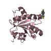
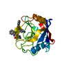
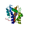
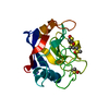
| Entry | Database: PDB / ID: 6gxg | ||||||
|---|---|---|---|---|---|---|---|
| Title | Tryparedoxin from Trypanosoma brucei in complex with CFT | ||||||
 Components Components | (Tryparedoxin) x 2 | ||||||
 Keywords Keywords | OXIDOREDUCTASE / Covalent Inhibitor / Photoreduction / Inhibitor-Induced Dimerization / Monomer-Dimer Mixture | ||||||
| Function / homology |  Function and homology information Function and homology informationthioredoxin-disulfide reductase (NADPH) activity / negative regulation of Wnt signaling pathway / negative regulation of protein ubiquitination / nucleus Similarity search - Function | ||||||
| Biological species |  | ||||||
| Method |  X-RAY DIFFRACTION / SYNCHROTRON / MOLECULAR REPLACEMENT / Resolution: 1.6 Å X-RAY DIFFRACTION / SYNCHROTRON / MOLECULAR REPLACEMENT / Resolution: 1.6 Å | ||||||
 Authors Authors | Bader, N. / Wagner, A. / Hellmich, U. / Schindelin, H. | ||||||
 Citation Citation | Journal: Angew Chem Int Ed Engl / Year: 2019 Title: Inhibitor-Induced Dimerization of an Essential Oxidoreductase from African Trypanosomes. Authors: Annika Wagner / Thien Anh Le / Martha Brennich / Philipp Klein / Nicole Bader / Erika Diehl / Daniel Paszek / A Katharina Weickhmann / Natalie Dirdjaja / R Luise Krauth-Siegel / Bernd Engels ...Authors: Annika Wagner / Thien Anh Le / Martha Brennich / Philipp Klein / Nicole Bader / Erika Diehl / Daniel Paszek / A Katharina Weickhmann / Natalie Dirdjaja / R Luise Krauth-Siegel / Bernd Engels / Till Opatz / Hermann Schindelin / Ute A Hellmich /   Abstract: Trypanosomal and leishmanial infections claim tens of thousands of lives each year. The metabolism of these unicellular eukaryotic parasites differs from the human host and their enzymes thus ...Trypanosomal and leishmanial infections claim tens of thousands of lives each year. The metabolism of these unicellular eukaryotic parasites differs from the human host and their enzymes thus constitute promising drug targets. Tryparedoxin (Tpx) from Trypanosoma brucei is the essential oxidoreductase in the parasite's hydroperoxide-clearance cascade. In vitro and in vivo functional assays show that a small, selective inhibitor efficiently inhibits Tpx. With X-ray crystallography, SAXS, analytical SEC, SEC-MALS, MD simulations, ITC, and NMR spectroscopy, we show how covalent binding of this monofunctional inhibitor leads to Tpx dimerization. Intra- and intermolecular inhibitor-inhibitor, protein-protein, and inhibitor-protein interactions stabilize the dimer. The behavior of this efficient antitrypanosomal molecule thus constitutes an exquisite example of chemically induced dimerization with a small, monovalent ligand that can be exploited for future drug design. | ||||||
| History |
|
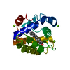
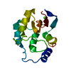
- Structure visualization
| Structure viewer | Molecule: MolmilJmol/JSmol |
|---|
- Downloads & links
-Download
| PDBx/mmCIF format | 6gxg.cif.gz | 112.8 KB | Display | PDBx/mmCIF format |
|---|---|---|---|---|
| PDB format | pdb6gxg.ent.gz | 86.9 KB | Display | PDB format |
| PDBx/mmJSON format | 6gxg.json.gz | Tree view | PDBx/mmJSON format | |
| Others |  Other downloads Other downloads |
-Validation report
| Arichive directory | https://data.pdbj.org/pub/pdb/validation_reports/gx/6gxgftp://data.pdbj.org/pub/pdb/validation_reports/gx/6gxg | HTTPS FTP |
|---|
-Related structure data
| Related structure data | 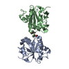 6gxyC 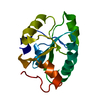 1o73S S: Starting model for refinement C: citing same article ( |
|---|---|
| Similar structure data |


-Links
 PDBj
PDBj

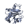
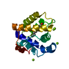
- Assembly
| Deposited unit | 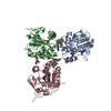
| ||||||||
|---|---|---|---|---|---|---|---|---|---|
| 1 | 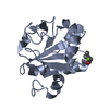
| ||||||||
| 2 | 
| ||||||||
| 3 | 
| ||||||||
| Unit cell |
|
-Components
| #1: Protein | Mass: 15878.901 Da / Num. of mol.: 1 Source method: isolated from a genetically manipulated source Source: (gene. exp.)  | ||||||
|---|---|---|---|---|---|---|---|
| #2: Protein | Mass: 16090.119 Da / Num. of mol.: 2 Source method: isolated from a genetically manipulated source Source: (gene. exp.) #3: Chemical | 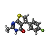  Mass: 260.287 Da / Num. of mol.: 3 / Source method: obtained synthetically / Formula: C13H9FN2OS / Feature type: SUBJECT OF INVESTIGATION Mass: 260.287 Da / Num. of mol.: 3 / Source method: obtained synthetically / Formula: C13H9FN2OS / Feature type: SUBJECT OF INVESTIGATION#4: Chemical |   Mass: 92.094 Da / Num. of mol.: 2 / Source method: obtained synthetically / Formula: C3H8O3 Mass: 92.094 Da / Num. of mol.: 2 / Source method: obtained synthetically / Formula: C3H8O3#5: Water | ChemComp-HOH / |  Mass: 18.015 Da / Num. of mol.: 418 / Source method: isolated from a natural source / Formula: H2O Mass: 18.015 Da / Num. of mol.: 418 / Source method: isolated from a natural source / Formula: H2O |
-Experimental details
-Experiment
| Experiment | Method: X-RAY DIFFRACTION / Number of used crystals: 1 |
|---|
- Sample preparation
| Crystal | Density Matthews: 1.87 Å3/Da / Density % sol: 34.3 % |
|---|---|
| Crystal grow | Temperature: 293 K / Method: vapor diffusion, sitting drop / pH: 5.5 / Details: 1.045 M Na-citrate; 100 mM Na-acetate buffer |
-Data collection
| Diffraction | Mean temperature: 100 K |
|---|---|
| Diffraction source | Source: SYNCHROTRON / Site: ESRF / Beamline: ID29 / Wavelength: 1.07227 Å |
| Detector | Type: DECTRIS PILATUS3 S 6M / Detector: PIXEL / Date: Jul 8, 2017 |
| Radiation | Protocol: SINGLE WAVELENGTH / Monochromatic (M) / Laue (L): M / Scattering type: x-ray |
| Radiation wavelength | Wavelength: 1.07227 Å / Relative weight: 1 |
| Reflection | Resolution: 1.6→50 Å / Num. obs: 45750 / % possible obs: 97.4 % / Redundancy: 6.8 % / Biso Wilson estimate: 11.9 Å2 / CC1/2: 0.996 / Rmerge(I) obs: 0.134 / Rpim(I) all: 0.056 / Rrim(I) all: 0.146 / Net I/σ(I): 9.6 |
| Reflection shell | Resolution: 1.6→1.63 Å / Redundancy: 7.1 % / Rmerge(I) obs: 1.441 / Num. unique obs: 2210 / CC1/2: 0.6 / Rpim(I) all: 0.577 / Rrim(I) all: 1.554 / % possible all: 95.3 |
- Processing
| Software |
| ||||||||||||||||||||||||||||||||||||||||||||||||||||||||||||||||||||||||||||||||||||||||||||||||||||||||||||||||||||||||||||||||||||||||||||||||||||||||||||||||||||||||||||||||||||||
|---|---|---|---|---|---|---|---|---|---|---|---|---|---|---|---|---|---|---|---|---|---|---|---|---|---|---|---|---|---|---|---|---|---|---|---|---|---|---|---|---|---|---|---|---|---|---|---|---|---|---|---|---|---|---|---|---|---|---|---|---|---|---|---|---|---|---|---|---|---|---|---|---|---|---|---|---|---|---|---|---|---|---|---|---|---|---|---|---|---|---|---|---|---|---|---|---|---|---|---|---|---|---|---|---|---|---|---|---|---|---|---|---|---|---|---|---|---|---|---|---|---|---|---|---|---|---|---|---|---|---|---|---|---|---|---|---|---|---|---|---|---|---|---|---|---|---|---|---|---|---|---|---|---|---|---|---|---|---|---|---|---|---|---|---|---|---|---|---|---|---|---|---|---|---|---|---|---|---|---|---|---|---|---|
| Refinement | Method to determine structure: MOLECULAR REPLACEMENT Starting model: 1o73 Resolution: 1.6→20 Å / Cor.coef. Fo:Fc: 0.974 / Cor.coef. Fo:Fc free: 0.962 / SU B: 2.017 / SU ML: 0.067 / Cross valid method: THROUGHOUT / ESU R: 0.095 / ESU R Free: 0.089 / Details: HYDROGENS HAVE BEEN ADDED IN THE RIDING POSITIONS
| ||||||||||||||||||||||||||||||||||||||||||||||||||||||||||||||||||||||||||||||||||||||||||||||||||||||||||||||||||||||||||||||||||||||||||||||||||||||||||||||||||||||||||||||||||||||
| Solvent computation | Ion probe radii: 0.8 Å / Shrinkage radii: 0.8 Å / VDW probe radii: 1.2 Å | ||||||||||||||||||||||||||||||||||||||||||||||||||||||||||||||||||||||||||||||||||||||||||||||||||||||||||||||||||||||||||||||||||||||||||||||||||||||||||||||||||||||||||||||||||||||
| Displacement parameters | Biso mean: 22.262 Å2
| ||||||||||||||||||||||||||||||||||||||||||||||||||||||||||||||||||||||||||||||||||||||||||||||||||||||||||||||||||||||||||||||||||||||||||||||||||||||||||||||||||||||||||||||||||||||
| Refinement step | Cycle: 1 / Resolution: 1.6→20 Å
| ||||||||||||||||||||||||||||||||||||||||||||||||||||||||||||||||||||||||||||||||||||||||||||||||||||||||||||||||||||||||||||||||||||||||||||||||||||||||||||||||||||||||||||||||||||||
| Refine LS restraints |
|