 Movie
Movie Controller

[English] 日本語
 Yorodumi
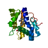
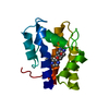
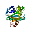
- PDB-4inx: Structure of Pheromone-binding protein 1 in complex with (Z,Z)-11... -
+ Open data

- Basic information
| Entry | Database: PDB / ID: 4inx | ||||||
|---|---|---|---|---|---|---|---|
| Title | Structure of Pheromone-binding protein 1 in complex with (Z,Z)-11,13- hexadecadienol | ||||||
 Components Components | Pheromone-binding protein 1 | ||||||
 Keywords Keywords | pheromone-binding protein / Amyelois transitella / pheromone / navel orangeworm moth / AtraPBP1 / pH-dependent binding | ||||||
| Function / homology |  Function and homology information Function and homology information | ||||||
| Biological species |  Amyelois transitella (butterflies/moths) Amyelois transitella (butterflies/moths) | ||||||
| Method |  X-RAY DIFFRACTION / SYNCHROTRON / MOLECULAR REPLACEMENT / molecular replacement / Resolution: 1.853 Å X-RAY DIFFRACTION / SYNCHROTRON / MOLECULAR REPLACEMENT / molecular replacement / Resolution: 1.853 Å | ||||||
 Authors Authors | di Luccio, E. / Wilson, D.K. | ||||||
 Citation Citation | Journal: Plos One / Year: 2013 Title: Crystallographic Observation of pH-Induced Conformational Changes in the Amyelois transitella Pheromone-Binding Protein AtraPBP1. Authors: di Luccio, E. / Ishida, Y. / Leal, W.S. / Wilson, D.K. | ||||||
| History |
|
- Structure visualization
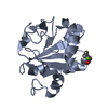
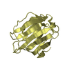
| Structure viewer | Molecule: MolmilJmol/JSmol |
|---|
- Downloads & links
-Download
| PDBx/mmCIF format | 4inx.cif.gz | 45.5 KB | Display | PDBx/mmCIF format |
|---|---|---|---|---|
| PDB format | pdb4inx.ent.gz | 31 KB | Display | PDB format |
| PDBx/mmJSON format | 4inx.json.gz | Tree view | PDBx/mmJSON format | |
| Others |  Other downloads Other downloads |
-Validation report
| Arichive directory | https://data.pdbj.org/pub/pdb/validation_reports/in/4inxftp://data.pdbj.org/pub/pdb/validation_reports/in/4inx | HTTPS FTP |
|---|
-Related structure data


-Links
 PDBj
PDBj- Assembly
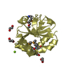
| Deposited unit | 
| ||||||||
|---|---|---|---|---|---|---|---|---|---|
| 1 |
| ||||||||
| Unit cell |
|
-Components
| #1: Protein | Mass: 15867.321 Da / Num. of mol.: 1 Source method: isolated from a genetically manipulated source Source: (gene. exp.) Amyelois transitella (butterflies/moths)Production host:  |
|---|---|
| #2: Chemical | ChemComp-1EX / (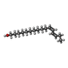  Mass: 238.409 Da / Num. of mol.: 1 / Source method: obtained synthetically / Formula: C16H30O Mass: 238.409 Da / Num. of mol.: 1 / Source method: obtained synthetically / Formula: C16H30O |
| #3: Water | ChemComp-HOH /  Mass: 18.015 Da / Num. of mol.: 161 / Source method: isolated from a natural source / Formula: H2O Mass: 18.015 Da / Num. of mol.: 161 / Source method: isolated from a natural source / Formula: H2O |
| Has protein modification | Y |
-Experimental details
-Experiment
| Experiment | Method: X-RAY DIFFRACTION / Number of used crystals: 1 |
|---|
- Sample preparation
| Crystal | Density Matthews: 2.82 Å3/Da / Density % sol: 56.31 % |
|---|---|
| Crystal grow | Temperature: 295 K / Method: vapor diffusion, hanging drop / pH: 6.5 Details: Purified AtraPBP1 was crystallized at room temperature by the hanging drop vapor diffusion method. Drops composed of 1ul protein solution (30 mg/ml) and 1ul of the precipitant solution were ...Details: Purified AtraPBP1 was crystallized at room temperature by the hanging drop vapor diffusion method. Drops composed of 1ul protein solution (30 mg/ml) and 1ul of the precipitant solution were suspended over a reservoir containing the precipitant solution (1.6 M sodium citrate pH 6.5). Crystals used in data collection were transferred into Paratone-N oil and flash-cooled in a stream of liquid nitrogen at 110 K, VAPOR DIFFUSION, HANGING DROP, temperature 295K |
-Data collection
| Diffraction | Mean temperature: 100 K | |||||||||||||||||||||||||||||||||||||||||||||||||||||||||||||||||||||||||||||
|---|---|---|---|---|---|---|---|---|---|---|---|---|---|---|---|---|---|---|---|---|---|---|---|---|---|---|---|---|---|---|---|---|---|---|---|---|---|---|---|---|---|---|---|---|---|---|---|---|---|---|---|---|---|---|---|---|---|---|---|---|---|---|---|---|---|---|---|---|---|---|---|---|---|---|---|---|---|---|
| Diffraction source | Source: SYNCHROTRON / Site: SSRL  / Beamline: BL9-2 / Wavelength: 1.2 Å / Beamline: BL9-2 / Wavelength: 1.2 Å | |||||||||||||||||||||||||||||||||||||||||||||||||||||||||||||||||||||||||||||
| Detector | Type: MARMOSAIC 325 mm CCD / Detector: CCD / Date: Apr 12, 2006 | |||||||||||||||||||||||||||||||||||||||||||||||||||||||||||||||||||||||||||||
| Radiation | Protocol: SINGLE WAVELENGTH / Monochromatic (M) / Laue (L): M / Scattering type: x-ray | |||||||||||||||||||||||||||||||||||||||||||||||||||||||||||||||||||||||||||||
| Radiation wavelength | Wavelength: 1.2 Å / Relative weight: 1 | |||||||||||||||||||||||||||||||||||||||||||||||||||||||||||||||||||||||||||||
| Reflection | Redundancy: 2.8 % / Av σ(I) over netI: 28.42 / Number: 41040 / Rmerge(I) obs: 0.04 / Χ2: 1.28 / D res high: 1.85 Å / D res low: 50 Å / Num. obs: 14800 / % possible obs: 98.3 | |||||||||||||||||||||||||||||||||||||||||||||||||||||||||||||||||||||||||||||
| Diffraction reflection shell |
| |||||||||||||||||||||||||||||||||||||||||||||||||||||||||||||||||||||||||||||
| Reflection | Resolution: 1.85→50 Å / Num. all: 15052 / Num. obs: 14800 / % possible obs: 98.3 % / Observed criterion σ(I): 28.4 / Redundancy: 2.8 % / Rmerge(I) obs: 0.04 / Χ2: 1.276 / Net I/σ(I): 16.3 | |||||||||||||||||||||||||||||||||||||||||||||||||||||||||||||||||||||||||||||
| Reflection shell |
|
-Phasing
| Phasing | Method: molecular replacement |
|---|
- Processing
| Software |
| |||||||||||||||||||||||||||||||||||||||||||||||||||||||||||||||||
|---|---|---|---|---|---|---|---|---|---|---|---|---|---|---|---|---|---|---|---|---|---|---|---|---|---|---|---|---|---|---|---|---|---|---|---|---|---|---|---|---|---|---|---|---|---|---|---|---|---|---|---|---|---|---|---|---|---|---|---|---|---|---|---|---|---|---|
| Refinement | Method to determine structure: MOLECULAR REPLACEMENT / Resolution: 1.853→34.1 Å / Cor.coef. Fo:Fc: 0.946 / Cor.coef. Fo:Fc free: 0.927 / Occupancy max: 1 / Occupancy min: 0.01 / SU B: 2.427 / SU ML: 0.076 / Cross valid method: THROUGHOUT / σ(F): 0 / ESU R: 0.121 / ESU R Free: 0.122 / Stereochemistry target values: MAXIMUM LIKELIHOOD Details: HYDROGENS HAVE BEEN ADDED IN THE RIDING POSITIONS U VALUES : REFINED INDIVIDUALLY
| |||||||||||||||||||||||||||||||||||||||||||||||||||||||||||||||||
| Solvent computation | Ion probe radii: 0.8 Å / Shrinkage radii: 0.8 Å / VDW probe radii: 1.4 Å / Solvent model: BABINET MODEL WITH MASK | |||||||||||||||||||||||||||||||||||||||||||||||||||||||||||||||||
| Displacement parameters | Biso max: 44.7 Å2 / Biso mean: 13.0968 Å2 / Biso min: 2.82 Å2
| |||||||||||||||||||||||||||||||||||||||||||||||||||||||||||||||||
| Refinement step | Cycle: LAST / Resolution: 1.853→34.1 Å
| |||||||||||||||||||||||||||||||||||||||||||||||||||||||||||||||||
| Refine LS restraints |
| |||||||||||||||||||||||||||||||||||||||||||||||||||||||||||||||||
| LS refinement shell | Resolution: 1.853→1.901 Å / Total num. of bins used: 20
|
