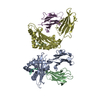
preprotein binding / cell envelope Sec protein transport complex / protein-exporting ATPase activity / protein-secreting ATPase / protein transport by the Sec complex / intracellular protein transmembrane transport / protein import / protein targeting to membrane / Secretion of toxins / protein secretion ...preprotein binding / cell envelope Sec protein transport complex / protein-exporting ATPase activity / protein-secreting ATPase / protein transport by the Sec complex / intracellular protein transmembrane transport / protein import / protein targeting to membrane / Secretion of toxins / protein secretion / ribonucleoprotein complex binding / protein targeting / cytoplasmic side of plasma membrane / protein transport / ribosome binding / zinc ion binding / ATP binding / identical protein binding / plasma membrane / cytoplasm Similarity search - Function
Protocol: SINGLE WAVELENGTH / Monochromatic (M) / Laue (L): M / Scattering type: x-ray
Radiation wavelength
Wavelength: 0.9795 Å / Relative weight: 1
Reflection
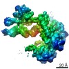
Resolution: 3.5→88.69 Å / Num. obs: 31379 / % possible obs: 99.4 % / Redundancy: 3.45 % / Net I/σ(I): 9.44
Reflection shell
Resolution: 3.5→3.71 Å
-
Processing
Software
Name
Version
Classification
XDS
datareduction
XSCALE
datascaling
REFMAC
5.8.0107
refinement
PDB_EXTRACT
3.24
dataextraction
PHASER
phasing
Refinement
Resolution: 3.5→88.69 Å / Cor.coef. Fo:Fc: 0.936 / Cor.coef. Fo:Fc free: 0.889 / SU B: 40.702 / SU ML: 0.607 / Cross valid method: THROUGHOUT / σ(F): 0 / ESU R Free: 0.662 Details: HYDROGENS HAVE BEEN ADDED IN THE RIDING POSITIONS U VALUES : REFINED INDIVIDUALLY
Rfactor
Num. reflection
% reflection
Selection details
Rfree
0.2927
785
4.9 %
RANDOM
Rwork
0.2315
-
-
-
obs
0.2344
15321
99.7 %
-
Solvent computation
Ion probe radii: 0.8 Å / Shrinkage radii: 0.8 Å / VDW probe radii: 1.2 Å
In the structure databanks used in Yorodumi, some data are registered as the other names, "COVID-19 virus" and "2019-nCoV". Here are the details of the virus and the list of structure data.
Jan 31, 2019. EMDB accession codes are about to change! (news from PDBe EMDB page)
EMDB accession codes are about to change! (news from PDBe EMDB page)
The allocation of 4 digits for EMDB accession codes will soon come to an end. Whilst these codes will remain in use, new EMDB accession codes will include an additional digit and will expand incrementally as the available range of codes is exhausted. The current 4-digit format prefixed with “EMD-” (i.e. EMD-XXXX) will advance to a 5-digit format (i.e. EMD-XXXXX), and so on. It is currently estimated that the 4-digit codes will be depleted around Spring 2019, at which point the 5-digit format will come into force.
The EM Navigator/Yorodumi systems omit the EMD- prefix.
Related info.:Q: What is EMD? / ID/Accession-code notation in Yorodumi/EM Navigator
Yorodumi is a browser for structure data from EMDB, PDB, SASBDB, etc.
This page is also the successor to EM Navigator detail page, and also detail information page/front-end page for Omokage search.
The word "yorodu" (or yorozu) is an old Japanese word meaning "ten thousand". "mi" (miru) is to see.
Related info.:EMDB / PDB / SASBDB / Comparison of 3 databanks / Yorodumi Search / Aug 31, 2016. New EM Navigator & Yorodumi / Yorodumi Papers / Jmol/JSmol / Function and homology information / Changes in new EM Navigator and Yorodumi
 Movie
Movie Controller


 Open data
 Basic information
 Components
 Keywords
 Function and homology information
 X-RAY DIFFRACTION /
  Authors
 Citation
 Structure visualization
 Downloads & links
 Other downloads


 PDBj
PDBj Assembly
 Sample preparation
 / Beamline: I02 / Wavelength: 0.9795 Å
 Processing