 Movie
Movie Controller


+ Open data

- Basic information
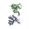
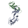
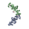
| Entry | Database: PDB / ID: 6gmm | ||||||
|---|---|---|---|---|---|---|---|
| Title | Crystal structure of Helicobacter pylori adhesin LabA | ||||||
 Components Components | adhesin LabA | ||||||
 Keywords Keywords | SUGAR BINDING PROTEIN / bacterial adhesin / lectin / Helicobacter pylori / gastric mucosa | ||||||
| Function / homology | SabA, N-terminal extracellular adhesion domain / SabA N-terminal extracellular adhesion domain / Outer membrane protein, Helicobacter / Helicobacter outer membrane protein / SabA N-terminal extracellular adhesion domain-containing protein Function and homology information Function and homology information | ||||||
| Biological species |   Helicobacter pylori (bacteria) Helicobacter pylori (bacteria) | ||||||
| Method |  X-RAY DIFFRACTION / SYNCHROTRON / MOLECULAR REPLACEMENT / Resolution: 2.06 Å X-RAY DIFFRACTION / SYNCHROTRON / MOLECULAR REPLACEMENT / Resolution: 2.06 Å | ||||||
 Authors Authors | Paraskevopoulou, V. / Overman, R.C. / Stolnik, S. / Winkler, S. / Gellert, P. / Falcone, F.H. / Schimpl, M. | ||||||
| Funding support |  United Kingdom, 1items United Kingdom, 1items
| ||||||
 Citation Citation | Journal: Current Research in Structural Biology / Year: 2021 Title: Structural and binding characterization of the LacdiNAc-specific adhesin (LabA; HopD) exodomain from Helicobacter pylori Authors: Paraskevopoulou, V. / Schimpl, M. / Overman, R.C. / Stolnik, S. / Chen, Y. / Nguyen, L. / Winkler, G.S. / Gellert, P. / Klassen, J.S. / Falcone, F.H. | ||||||
| History |
|
- Structure visualization
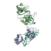
| Structure viewer | Molecule: MolmilJmol/JSmol |
|---|
- Downloads & links
-Download
| PDBx/mmCIF format | 6gmm.cif.gz | 178.7 KB | Display | PDBx/mmCIF format |
|---|---|---|---|---|
| PDB format | pdb6gmm.ent.gz | 139.1 KB | Display | PDB format |
| PDBx/mmJSON format | 6gmm.json.gz | Tree view | PDBx/mmJSON format | |
| Others |  Other downloads Other downloads |
-Validation report
| Arichive directory | https://data.pdbj.org/pub/pdb/validation_reports/gm/6gmmftp://data.pdbj.org/pub/pdb/validation_reports/gm/6gmm | HTTPS FTP |
|---|
-Related structure data
| Related structure data | 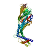 4zh7S S: Starting model for refinement |
|---|---|
| Similar structure data |


-Links
 PDBj
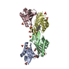
PDBj- Assembly
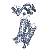
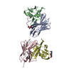
| Deposited unit | 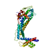
| ||||||||
|---|---|---|---|---|---|---|---|---|---|
| 1 |
| ||||||||
| Unit cell |
|
-Components
| #1: Protein | Mass: 48788.566 Da / Num. of mol.: 1 Source method: isolated from a genetically manipulated source Source: (gene. exp.) Helicobacter pylori (bacteria) / Gene: BW246_00440, BZL55_00170 / Production host: |
|---|---|
| #2: Water | ChemComp-HOH /  Mass: 18.015 Da / Num. of mol.: 173 / Source method: isolated from a natural source / Formula: H2O Mass: 18.015 Da / Num. of mol.: 173 / Source method: isolated from a natural source / Formula: H2O |
| Has protein modification | Y |
-Experimental details
-Experiment
| Experiment | Method: X-RAY DIFFRACTION / Number of used crystals: 1 |
|---|
- Sample preparation
| Crystal | Density Matthews: 2.54 Å3/Da / Density % sol: 51.59 % / Description: bipyramid |
|---|---|
| Crystal grow | Temperature: 293 K / Method: vapor diffusion, sitting drop / pH: 6.5 / Details: 28 % PEG 2000 MME, 0.1 M Bis-Tris pH 6.5 |
-Data collection
| Diffraction | Mean temperature: 100 K / Serial crystal experiment: N |
|---|---|
| Diffraction source | Source: SYNCHROTRON / Site: Diamond / Beamline: I04 / Wavelength: 0.9795 Å |
| Detector | Type: DECTRIS PILATUS3 6M / Detector: PIXEL / Date: Feb 9, 2018 |
| Radiation | Protocol: SINGLE WAVELENGTH / Monochromatic (M) / Laue (L): M / Scattering type: x-ray |
| Radiation wavelength | Wavelength: 0.9795 Å / Relative weight: 1 |
| Reflection | Resolution: 2.056→49.537 Å / Num. obs: 31169 / % possible obs: 99.9 % / Redundancy: 12.3 % / Biso Wilson estimate: 20.37 Å2 / CC1/2: 0.999 / Rmerge(I) obs: 0.242 / Net I/σ(I): 8.1 |
| Reflection shell | Resolution: 2.056→2.093 Å / Redundancy: 11 % / Rmerge(I) obs: 1.516 / Mean I/σ(I) obs: 1.9 / Num. unique obs: 1558 / CC1/2: 0.599 / % possible all: 98.9 |
- Processing
| Software |
| ||||||||||||||||||||||||||||||||||||||||||||||||||||||||||||||||||||||||||||||||||||||||||||||||||||||||||||||||||
|---|---|---|---|---|---|---|---|---|---|---|---|---|---|---|---|---|---|---|---|---|---|---|---|---|---|---|---|---|---|---|---|---|---|---|---|---|---|---|---|---|---|---|---|---|---|---|---|---|---|---|---|---|---|---|---|---|---|---|---|---|---|---|---|---|---|---|---|---|---|---|---|---|---|---|---|---|---|---|---|---|---|---|---|---|---|---|---|---|---|---|---|---|---|---|---|---|---|---|---|---|---|---|---|---|---|---|---|---|---|---|---|---|---|---|---|
| Refinement | Method to determine structure: MOLECULAR REPLACEMENT Starting model: 4ZH7 Resolution: 2.06→49.537 Å / Cor.coef. Fo:Fc: 0.914 / Cor.coef. Fo:Fc free: 0.889 / Rfactor Rfree error: 0 / SU R Cruickshank DPI: 0.187 / Cross valid method: THROUGHOUT / σ(F): 0 / SU R Blow DPI: 0.197 / SU Rfree Blow DPI: 0.176 / SU Rfree Cruickshank DPI: 0.172
| ||||||||||||||||||||||||||||||||||||||||||||||||||||||||||||||||||||||||||||||||||||||||||||||||||||||||||||||||||
| Displacement parameters | Biso mean: 36.27 Å2
| ||||||||||||||||||||||||||||||||||||||||||||||||||||||||||||||||||||||||||||||||||||||||||||||||||||||||||||||||||
| Refine analyze | Luzzati coordinate error obs: 0.34 Å | ||||||||||||||||||||||||||||||||||||||||||||||||||||||||||||||||||||||||||||||||||||||||||||||||||||||||||||||||||
| Refinement step | Cycle: 1 / Resolution: 2.06→49.537 Å
| ||||||||||||||||||||||||||||||||||||||||||||||||||||||||||||||||||||||||||||||||||||||||||||||||||||||||||||||||||
| Refine LS restraints |
| ||||||||||||||||||||||||||||||||||||||||||||||||||||||||||||||||||||||||||||||||||||||||||||||||||||||||||||||||||
| LS refinement shell | Resolution: 2.06→2.13 Å / Rfactor Rfree error: 0 / Total num. of bins used: 16
| ||||||||||||||||||||||||||||||||||||||||||||||||||||||||||||||||||||||||||||||||||||||||||||||||||||||||||||||||||
| Refinement TLS params. | Method: refined / Origin x: -5.2603 Å / Origin y: 7.8779 Å / Origin z: -24.0345 Å
| ||||||||||||||||||||||||||||||||||||||||||||||||||||||||||||||||||||||||||||||||||||||||||||||||||||||||||||||||||
| Refinement TLS group | Selection details: { A|* } |
