 Movie
Movie Controller

[English] 日本語
 Yorodumi
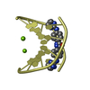
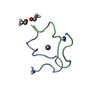
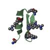
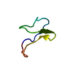
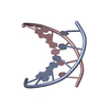
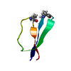
- PDB-6gim: Structure of the DNA duplex d(AAATTT)2 with [N-(3-chloro-4-((4,5-... -
+ Open data

- Basic information
| Entry | Database: PDB / ID: 6gim | |||||||||||||||||||||||||||||||||||||
|---|---|---|---|---|---|---|---|---|---|---|---|---|---|---|---|---|---|---|---|---|---|---|---|---|---|---|---|---|---|---|---|---|---|---|---|---|---|---|
| Title | Structure of the DNA duplex d(AAATTT)2 with [N-(3-chloro-4-((4,5-dihydro-1H-imidazol-2-yl)amino)phenyl)-4-((4,5-dihydro-1H-imidazol-2- yl)amino)benzamide] - (drug JNI18) | |||||||||||||||||||||||||||||||||||||
 Components Components | DNA (5'-D(* Keywords KeywordsDNA / AT-rich DNA / DNA binding drugs / Minor groove binding drugs / antiparasitic drugs / Trypanosoma brucei | Function / homology | Chem-EZK / DNA |  Function and homology information Function and homology informationBiological species |  Method |  X-RAY DIFFRACTION / SYNCHROTRON / MOLECULAR REPLACEMENT / Resolution: 1.43 Å X-RAY DIFFRACTION / SYNCHROTRON / MOLECULAR REPLACEMENT / Resolution: 1.43 Å  Authors AuthorsMillan, C.R. / Dardonvile, C. / de Koning, H.P. / Saperas, N. / Campos, J.L. | Funding support | |  Spain, Spain,  United Kingdom, 4items United Kingdom, 4items
 CitationJournal: Nucleic Acids Res. / Year: 2017 CitationJournal: Nucleic Acids Res. / Year: 2017Title: Functional and structural analysis of AT-specific minor groove binders that disrupt DNA-protein interactions and cause disintegration of the Trypanosoma brucei kinetoplast. Authors: Millan, C.R. / Acosta-Reyes, F.J. / Lagartera, L. / Ebiloma, G.U. / Lemgruber, L. / Nue Martinez, J.J. / Saperas, N. / Dardonville, C. / de Koning, H.P. / Campos, J.L. History |
|
- Structure visualization
| Structure viewer | Molecule: MolmilJmol/JSmol |
|---|
- Downloads & links
-Download
| PDBx/mmCIF format | 6gim.cif.gz | 46 KB | Display | PDBx/mmCIF format |
|---|---|---|---|---|
| PDB format | pdb6gim.ent.gz | 33.3 KB | Display | PDB format |
| PDBx/mmJSON format | 6gim.json.gz | Tree view | PDBx/mmJSON format | |
| Others |  Other downloads Other downloads |
-Validation report
| Arichive directory | https://data.pdbj.org/pub/pdb/validation_reports/gi/6gimftp://data.pdbj.org/pub/pdb/validation_reports/gi/6gim | HTTPS FTP |
|---|
-Related structure data


-Links
 PDBj
PDBj

- Assembly
| Deposited unit | 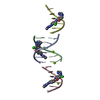
| ||||||||||||
|---|---|---|---|---|---|---|---|---|---|---|---|---|---|
| 1 | 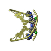
| ||||||||||||
| 2 | 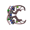
| ||||||||||||
| 3 | 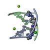
| ||||||||||||
| Unit cell |
| ||||||||||||
| Components on special symmetry positions |
|
-Components
| #1: DNA chain | Mass: 1807.241 Da / Num. of mol.: 4 / Source method: obtained synthetically / Source: (synth.) #2: Chemical | 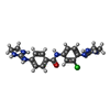  Mass: 399.877 Da / Num. of mol.: 3 / Source method: obtained synthetically / Formula: C19H22ClN7O Mass: 399.877 Da / Num. of mol.: 3 / Source method: obtained synthetically / Formula: C19H22ClN7O#3: Chemical |   Mass: 24.305 Da / Num. of mol.: 3 / Source method: obtained synthetically / Formula: Mg Mass: 24.305 Da / Num. of mol.: 3 / Source method: obtained synthetically / Formula: Mg#4: Water | ChemComp-HOH / |  Mass: 18.015 Da / Num. of mol.: 95 / Source method: isolated from a natural source / Formula: H2O Mass: 18.015 Da / Num. of mol.: 95 / Source method: isolated from a natural source / Formula: H2O |
|---|
-Experimental details
-Experiment
| Experiment | Method: X-RAY DIFFRACTION / Number of used crystals: 1 |
|---|
- Sample preparation
| Crystal | Density Matthews: 2.304846 Å3/Da / Density meas: 0.127 Mg/m3 / Density % sol: 46.66 % |
|---|---|
| Crystal grow | Temperature: 277 K / Method: vapor diffusion, hanging drop / pH: 6 Details: 10 mM magnesium acetate, 0.1 mM spermine, 5% 2-methyl-2,4-pentanediol (MPD); equilibrated against 20% MPD reservoir |
-Data collection
| Diffraction | Mean temperature: 100 K |
|---|---|
| Diffraction source | Source: SYNCHROTRON / Site: ALBA / Beamline: XALOC / Wavelength: 0.9791 Å |
| Detector | Type: DECTRIS PILATUS 6M / Detector: PIXEL / Date: Jul 27, 2017 |
| Radiation | Protocol: SINGLE WAVELENGTH / Monochromatic (M) / Laue (L): M / Scattering type: x-ray |
| Radiation wavelength | Wavelength: 0.9791 Å / Relative weight: 1 |
| Reflection | Resolution: 1.43→36.04 Å / Num. obs: 12256 / % possible obs: 99.5 % / Redundancy: 4 % / Biso Wilson estimate: 12.7 Å2 / CC1/2: 0.995 / Rmerge(I) obs: 0.073 / Rpim(I) all: 0.033 / Rrim(I) all: 0.081 / Χ2: 0.92 / Net I/av σ(I): 13.4 / Net I/σ(I): 2.79 |
| Reflection shell | Resolution: 1.43→7.81 Å / Rmerge(I) obs: 0.581 / Mean I/σ(I) obs: 3.1 / Num. unique obs: 602 / CC1/2: 0.858 / Rpim(I) all: 0.376 / Rrim(I) all: 0.642 / Χ2: 0.72 / % possible all: 99.7 |
- Processing
| Software |
| ||||||||||||||||||||||||||||||||||||||||||||||||||||||||||||||||||||||||||||||||||||||||||||||||||||||||||||||||||||||||||||||||||||||||||||||||||||||||||||||||||||||||||||||||||||||
|---|---|---|---|---|---|---|---|---|---|---|---|---|---|---|---|---|---|---|---|---|---|---|---|---|---|---|---|---|---|---|---|---|---|---|---|---|---|---|---|---|---|---|---|---|---|---|---|---|---|---|---|---|---|---|---|---|---|---|---|---|---|---|---|---|---|---|---|---|---|---|---|---|---|---|---|---|---|---|---|---|---|---|---|---|---|---|---|---|---|---|---|---|---|---|---|---|---|---|---|---|---|---|---|---|---|---|---|---|---|---|---|---|---|---|---|---|---|---|---|---|---|---|---|---|---|---|---|---|---|---|---|---|---|---|---|---|---|---|---|---|---|---|---|---|---|---|---|---|---|---|---|---|---|---|---|---|---|---|---|---|---|---|---|---|---|---|---|---|---|---|---|---|---|---|---|---|---|---|---|---|---|---|---|
| Refinement | Method to determine structure: MOLECULAR REPLACEMENT Starting model: Idealised DNA model from TURBO Resolution: 1.43→36.04 Å / Cor.coef. Fo:Fc: 0.978 / Cor.coef. Fo:Fc free: 0.948 / SU B: 2.476 / SU ML: 0.044 / Cross valid method: THROUGHOUT / ESU R: 0.074 / ESU R Free: 0.068 / Details: HYDROGENS HAVE BEEN ADDED IN THE RIDING POSITIONS
| ||||||||||||||||||||||||||||||||||||||||||||||||||||||||||||||||||||||||||||||||||||||||||||||||||||||||||||||||||||||||||||||||||||||||||||||||||||||||||||||||||||||||||||||||||||||
| Solvent computation | Ion probe radii: 0.8 Å / Shrinkage radii: 0.8 Å / VDW probe radii: 1.2 Å | ||||||||||||||||||||||||||||||||||||||||||||||||||||||||||||||||||||||||||||||||||||||||||||||||||||||||||||||||||||||||||||||||||||||||||||||||||||||||||||||||||||||||||||||||||||||
| Displacement parameters | Biso mean: 16.164 Å2
| ||||||||||||||||||||||||||||||||||||||||||||||||||||||||||||||||||||||||||||||||||||||||||||||||||||||||||||||||||||||||||||||||||||||||||||||||||||||||||||||||||||||||||||||||||||||
| Refinement step | Cycle: 1 / Resolution: 1.43→36.04 Å
| ||||||||||||||||||||||||||||||||||||||||||||||||||||||||||||||||||||||||||||||||||||||||||||||||||||||||||||||||||||||||||||||||||||||||||||||||||||||||||||||||||||||||||||||||||||||
| Refine LS restraints |
|