 Movie
Movie Controller


+ Open data

- Basic information
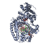
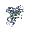
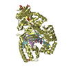
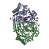
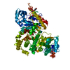
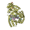
| Entry | Database: PDB / ID: 6ghs | ||||||
|---|---|---|---|---|---|---|---|
| Title | Modification dependent TagI restriction endonuclease | ||||||
 Components Components | TagI restriction endonuclease | ||||||
 Keywords Keywords | HYDROLASE / RESTRICTION ENDONUCLEASE / TYPE II / TYPE IV / CYTOSINE MODIFICATION / 5-METHYLCYTOSINE / 5MC / 5-HYDROXYMETHYLCYTOSINE / 5HMC / SRA / HNH / BBA-ME NUCLEASE / ScoA3IV / SCO5333 / TbiR51I / TBIS1 | ||||||
| Function / homology |  Function and homology information Function and homology informationubiquitin protein ligase activity / protein ubiquitination / metal ion binding Similarity search - Function | ||||||
| Biological species |  Thermocrispum agreste (bacteria) Thermocrispum agreste (bacteria) | ||||||
| Method |  X-RAY DIFFRACTION / MOLECULAR REPLACEMENT / Resolution: 2.92 Å X-RAY DIFFRACTION / MOLECULAR REPLACEMENT / Resolution: 2.92 Å | ||||||
 Authors Authors | Kisiala, M. / Copelas, A. / Czapinska, H. / Xu, S. / Bochtler, M. | ||||||
 Citation Citation | Journal: Nucleic Acids Res. / Year: 2018 Title: Crystal structure of the modification-dependent SRA-HNH endonuclease TagI. Authors: Kisiala, M. / Copelas, A. / Czapinska, H. / Xu, S.Y. / Bochtler, M. | ||||||
| History |
|
- Structure visualization
| Structure viewer | Molecule: MolmilJmol/JSmol |
|---|
- Downloads & links
-Download
| PDBx/mmCIF format | 6ghs.cif.gz | 71.6 KB | Display | PDBx/mmCIF format |
|---|---|---|---|---|
| PDB format | pdb6ghs.ent.gz | 51.7 KB | Display | PDB format |
| PDBx/mmJSON format | 6ghs.json.gz | Tree view | PDBx/mmJSON format | |
| Others |  Other downloads Other downloads |
-Validation report
| Summary document | 6ghs_validation.pdf.gz | 416.1 KB | Display | wwPDB validaton report |
|---|---|---|---|---|
| Full document | 6ghs_full_validation.pdf.gz | 416.1 KB | Display | |
| Data in XML | 6ghs_validation.xml.gz | 12 KB | Display | |
| Data in CIF | 6ghs_validation.cif.gz | 16.1 KB | Display | |
| Arichive directory | https://data.pdbj.org/pub/pdb/validation_reports/gh/6ghsftp://data.pdbj.org/pub/pdb/validation_reports/gh/6ghs | HTTPS FTP |
-Related structure data


-Links
 PDBj
PDBj

- Assembly
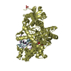
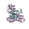
| Deposited unit | 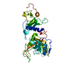
| ||||||||
|---|---|---|---|---|---|---|---|---|---|
| 1 | 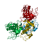
| ||||||||
| Unit cell |
|
-Components
| #1: Protein | Mass: 33997.176 Da / Num. of mol.: 1 Source method: isolated from a genetically manipulated source Source: (gene. exp.) Thermocrispum agreste (bacteria) / Plasmid: pTXB1 / Production host: |
|---|---|
| #2: Chemical | ChemComp-ZN /   Mass: 65.409 Da / Num. of mol.: 1 / Source method: obtained synthetically / Formula: Zn Mass: 65.409 Da / Num. of mol.: 1 / Source method: obtained synthetically / Formula: Zn |
| #3: Chemical | ChemComp-NA /   Mass: 22.990 Da / Num. of mol.: 1 / Source method: obtained synthetically / Formula: Na Mass: 22.990 Da / Num. of mol.: 1 / Source method: obtained synthetically / Formula: Na |
| #4: Water | ChemComp-HOH /  Mass: 18.015 Da / Num. of mol.: 53 / Source method: isolated from a natural source / Formula: H2O Mass: 18.015 Da / Num. of mol.: 53 / Source method: isolated from a natural source / Formula: H2O |
-Experimental details
-Experiment
| Experiment | Method: X-RAY DIFFRACTION / Number of used crystals: 1 |
|---|
- Sample preparation
| Crystal | Density Matthews: 4.4 Å3/Da / Density % sol: 72.07 % |
|---|---|
| Crystal grow | Temperature: 294 K / Method: liquid diffusion / pH: 7.5 Details: A5 Morpheus Buffer (10% w/v PEG 20 000, 20% v/v PEG MME 550, 0.03 M MgCl2, 0.03 M CaCl2, 0.1 M MOPS/HEPES-Na pH 7.5) PH range: 7.5 |
-Data collection
| Diffraction | Mean temperature: 294 K |
|---|---|
| Diffraction source | Source: ROTATING ANODE / Type: BRUKER X8 PROTEUM / Wavelength: 1.54 Å |
| Detector | Type: Bruker Platinum 135 / Detector: CCD / Date: Feb 16, 2018 / Details: HELIOS multilayer optics |
| Radiation | Protocol: SINGLE WAVELENGTH / Monochromatic (M) / Laue (L): M / Scattering type: x-ray |
| Radiation wavelength | Wavelength: 1.54 Å / Relative weight: 1 |
| Reflection | Resolution: 2.92→20 Å / Num. obs: 12496 / % possible obs: 96.1 % / Redundancy: 3.23 % / Rmerge(I) obs: 0.2386 / Rsym value: 0.2196 / Net I/σ(I): 4.38 |
| Reflection shell | Resolution: 2.92→3.02 Å / Redundancy: 3.3 % / Rmerge(I) obs: 0.6983 / Mean I/σ(I) obs: 1.2 / Rsym value: 0.9485 / % possible all: 96.2 |
- Processing
| Software |
| ||||||||||||||||||||||||||||||||||||||||||||||||||||||||||||||||||||||||||||||||||||||||||||||||||||||||||||||||||||||||||||||||||||||||||||||||||||||||||||||||||||||||||||||||||||||
|---|---|---|---|---|---|---|---|---|---|---|---|---|---|---|---|---|---|---|---|---|---|---|---|---|---|---|---|---|---|---|---|---|---|---|---|---|---|---|---|---|---|---|---|---|---|---|---|---|---|---|---|---|---|---|---|---|---|---|---|---|---|---|---|---|---|---|---|---|---|---|---|---|---|---|---|---|---|---|---|---|---|---|---|---|---|---|---|---|---|---|---|---|---|---|---|---|---|---|---|---|---|---|---|---|---|---|---|---|---|---|---|---|---|---|---|---|---|---|---|---|---|---|---|---|---|---|---|---|---|---|---|---|---|---|---|---|---|---|---|---|---|---|---|---|---|---|---|---|---|---|---|---|---|---|---|---|---|---|---|---|---|---|---|---|---|---|---|---|---|---|---|---|---|---|---|---|---|---|---|---|---|---|---|
| Refinement | Method to determine structure: MOLECULAR REPLACEMENT Starting model: 3clz, 5mkw Resolution: 2.92→20 Å / Cor.coef. Fo:Fc: 0.893 / Cor.coef. Fo:Fc free: 0.911 / SU B: 16.582 / SU ML: 0.279 / Cross valid method: THROUGHOUT / ESU R: 0.523 / ESU R Free: 0.293 Details: THE IDENTITY OF THE METAL ION IN THE ACTIVE SITE (LIGATED BY HIS 254 AND ASP 258) IS UNCERTAIN. UNDER PHYSIOLOGICAL CONDITIONS THIS SITE IS MOST LIKELY OCCUPIED BY A MG2+ ION. HOWEVER, SINCE ...Details: THE IDENTITY OF THE METAL ION IN THE ACTIVE SITE (LIGATED BY HIS 254 AND ASP 258) IS UNCERTAIN. UNDER PHYSIOLOGICAL CONDITIONS THIS SITE IS MOST LIKELY OCCUPIED BY A MG2+ ION. HOWEVER, SINCE THE CONCENTRATION OF EDTA IN THE PROTEIN BUFFER EXCEEDS THE MG2+ CONCENTRATION IN THE RESERVOIR BUFFER ALMOST 20 TIMES WE PREDICT THAT THE MG2+ IONS (AND CA2+ IONS THAT WERE ALSO PRESENT IN THE BUFFER) WERE DEPLETED. UNFORTUNATELY, THE RESOLUTION OF THE DIFFRACTION DATA IS NOT HIGH ENOUGH TO PERFORM THE ANALYSIS OF THE METAL-LIGAND DISTANCES, BUT SINCE SODIUM IONS WERE ABUNDANT IN BOTH BUFFERS AND SINCE THE ELECTRON DENSITY LEVEL OF THE ION ROUGHLY CORRESPONDS TO ONE OF THE SURROUNDING PROTEIN AND SOLVENT ATOMS, WE HAVE TENTATIVELY ASSIGNED THE METAL IN THE ACTIVE SITE AS A NA+ ION. HYDROGENS HAVE BEEN ADDED IN THE RIDING POSITIONS
| ||||||||||||||||||||||||||||||||||||||||||||||||||||||||||||||||||||||||||||||||||||||||||||||||||||||||||||||||||||||||||||||||||||||||||||||||||||||||||||||||||||||||||||||||||||||
| Solvent computation | Ion probe radii: 0.8 Å / Shrinkage radii: 0.8 Å / VDW probe radii: 1.4 Å | ||||||||||||||||||||||||||||||||||||||||||||||||||||||||||||||||||||||||||||||||||||||||||||||||||||||||||||||||||||||||||||||||||||||||||||||||||||||||||||||||||||||||||||||||||||||
| Displacement parameters | Biso mean: 51.062 Å2
| ||||||||||||||||||||||||||||||||||||||||||||||||||||||||||||||||||||||||||||||||||||||||||||||||||||||||||||||||||||||||||||||||||||||||||||||||||||||||||||||||||||||||||||||||||||||
| Refinement step | Cycle: LAST / Resolution: 2.92→20 Å
| ||||||||||||||||||||||||||||||||||||||||||||||||||||||||||||||||||||||||||||||||||||||||||||||||||||||||||||||||||||||||||||||||||||||||||||||||||||||||||||||||||||||||||||||||||||||
| Refine LS restraints |
|