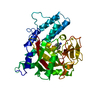
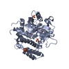
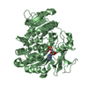
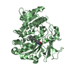
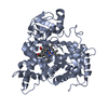
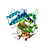
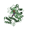
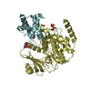
Entry Database : PDB / ID : 6ghkTitle Human PARP1 (ARTD1) - Catalytic domain in complex with inhibitor ME0527 Poly [ADP-ribose] polymerase 1 Keywords / / / Function / homology Function Domain/homology Component
/ / / / / / / / / / / / / / / / / / / / / / / / / / / / / / / / / / / / / / / / / / / / / / / / / / / / / / / / / / / / / / / / / / / / / / / / / / / / / / / / / / / / / / / / / / / / / / / / / / / / / / / / / / / / / / / / / / / / / / / / / / / / / / / / / / / / / / / / / / / / / / / / / / / / / / / / Biological species Homo sapiens (human)Method / / / Resolution : 2.28 Å Authors Karlberg, T. / Thorsell, A.G. / Lindgren, A.E.G. / Moche, M. / Brock, J. / Ekblad, T. / Spjut, S. / Elofsson, M. / Schuler, H. Journal : To Be Published Title : Human PARP1 (ARTD1) - Catalytic domain in complex with inhibitor ME0527Authors : Karlberg, T. / Thorsell, A.G. / Lindgren, A.E.G. / Ekblad, T. / Spjut, S. / Elofsson, M. / Schuler, H. History Deposition May 8, 2018 Deposition site / Processing site Revision 1.0 May 22, 2019 Provider / Type Revision 1.1 Jan 17, 2024 Group / Database references / Refinement descriptionCategory chem_comp_atom / chem_comp_bond ... chem_comp_atom / chem_comp_bond / database_2 / pdbx_initial_refinement_model Item / _database_2.pdbx_database_accession
Show all Show less
 Movie
Movie Controller

 Yorodumi
 Open data
 Basic information
 Components
 Keywords
 Function and homology information
 Homo sapiens (human)
 X-RAY DIFFRACTION /
  Authors
 Citation
 Structure visualization
 Downloads & links
 Other downloads
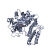


 PDBj
PDBj


 Assembly
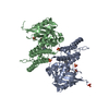
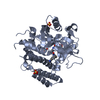
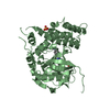


 Mass: 387.434 Da / Num. of mol.: 2 / Source method: obtained synthetically / Formula: C22H21N5O2
 Mass: 96.063 Da / Num. of mol.: 6 / Source method: obtained synthetically / Formula: SO4
 Mass: 18.015 Da / Num. of mol.: 208 / Source method: isolated from a natural source / Formula: H2O
 Sample preparation
 / Beamline: 14.1 / Wavelength: 0.91841 Å
 Processing