 Movie
Movie Controller

[English] 日本語
 Yorodumi
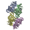
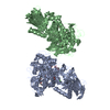
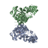
- PDB-6gcu: MET receptor in complex with InlB internalin domain and DARPin A3A -
+ Open data

- Basic information
| Entry | Database: PDB / ID: 6gcu | ||||||
|---|---|---|---|---|---|---|---|
| Title | MET receptor in complex with InlB internalin domain and DARPin A3A | ||||||
 Components Components |
| ||||||
 Keywords Keywords | SIGNALING PROTEIN / receptor tyrosine kinase / bacterial invasion protein / artificial binding protein | ||||||
| Function / homology |  Function and homology information Function and homology informationentry of bacterium into host cell / hepatocyte growth factor receptor activity / Drug-mediated inhibition of MET activation / MET activates STAT3 / negative regulation of hydrogen peroxide-mediated programmed cell death / MET Receptor Activation / MET interacts with TNS proteins / endothelial cell morphogenesis / semaphorin receptor activity / MET receptor recycling ...entry of bacterium into host cell / hepatocyte growth factor receptor activity / Drug-mediated inhibition of MET activation / MET activates STAT3 / negative regulation of hydrogen peroxide-mediated programmed cell death / MET Receptor Activation / MET interacts with TNS proteins / endothelial cell morphogenesis / semaphorin receptor activity / MET receptor recycling / pancreas development / MET activates PTPN11 / hepatocyte growth factor receptor signaling pathway / MET activates RAP1 and RAC1 / Sema4D mediated inhibition of cell attachment and migration / positive regulation of endothelial cell chemotaxis / MET activates PI3K/AKT signaling / MET activates PTK2 signaling / branching morphogenesis of an epithelial tube / positive chemotaxis / semaphorin-plexin signaling pathway / Regulation of MITF-M-dependent genes involved in cell cycle and proliferation / MET activates RAS signaling / MECP2 regulates neuronal receptors and channels / peptidoglycan-based cell wall / cell surface receptor protein tyrosine kinase signaling pathway / basal plasma membrane / molecular function activator activity / excitatory postsynaptic potential / liver development / negative regulation of autophagy / InlB-mediated entry of Listeria monocytogenes into host cell / receptor protein-tyrosine kinase / Negative regulation of MET activity / neuron differentiation / Constitutive Signaling by Aberrant PI3K in Cancer / PIP3 activates AKT signaling / heparin binding / PI5P, PP2A and IER3 Regulate PI3K/AKT Signaling / RAF/MAP kinase cascade / protein tyrosine kinase activity / protein phosphatase binding / cell surface receptor signaling pathway / postsynapse / signaling receptor complex / lipid binding / cell surface / positive regulation of transcription by RNA polymerase II / extracellular region / ATP binding / membrane / metal ion binding / identical protein binding / plasma membrane Similarity search - Function | ||||||
| Biological species |  Homo sapiens (human) Homo sapiens (human) Listeria monocytogenes serovar 1/2a (bacteria) Listeria monocytogenes serovar 1/2a (bacteria)synthetic construct (others) | ||||||
| Method |  X-RAY DIFFRACTION / SYNCHROTRON / MOLECULAR REPLACEMENT / Resolution: 6.001 Å X-RAY DIFFRACTION / SYNCHROTRON / MOLECULAR REPLACEMENT / Resolution: 6.001 Å | ||||||
 Authors Authors | Meyer, T. / Andres, F. / Iamele, L. / Gherardi, E. / Pluckthun, A. / Niemann, H.H. | ||||||
| Funding support |  Germany, 1items Germany, 1items
| ||||||
 Citation Citation | Journal: J.Mol.Biol. / Year: 2019 Title: Inhibition of the MET Kinase Activity and Cell Growth in MET-Addicted Cancer Cells by Bi-Paratopic Linking. Authors: Andres, F. / Iamele, L. / Meyer, T. / Stuber, J.C. / Kast, F. / Gherardi, E. / Niemann, H.H. / Pluckthun, A. | ||||||
| History |
|
- Structure visualization
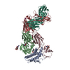
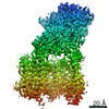
| Structure viewer | Molecule: MolmilJmol/JSmol |
|---|
- Downloads & links
-Download
| PDBx/mmCIF format | 6gcu.cif.gz | 442.8 KB | Display | PDBx/mmCIF format |
|---|---|---|---|---|
| PDB format | pdb6gcu.ent.gz | 352.7 KB | Display | PDB format |
| PDBx/mmJSON format | 6gcu.json.gz | Tree view | PDBx/mmJSON format | |
| Others |  Other downloads Other downloads |
-Validation report
| Arichive directory | https://data.pdbj.org/pub/pdb/validation_reports/gc/6gcuftp://data.pdbj.org/pub/pdb/validation_reports/gc/6gcu | HTTPS FTP |
|---|
-Related structure data
| Related structure data | 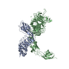 2uzyS S: Starting model for refinement |
|---|---|
| Similar structure data |

-Links
 PDBj
PDBj


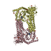
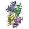
- Assembly
| Deposited unit | 
| ||||||||
|---|---|---|---|---|---|---|---|---|---|
| 1 | 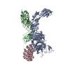
| ||||||||
| 2 | 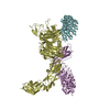
| ||||||||
| Unit cell |
|
-Components
| #1: Protein | Mass: 81949.047 Da / Num. of mol.: 2 Source method: isolated from a genetically manipulated source Source: (gene. exp.) Homo sapiens (human) / Gene: MET / Cell line (production host): CHO lec3.2.8.1 / Production host:   Cricetulus griseus (Chinese hamster) Cricetulus griseus (Chinese hamster)References: UniProt: P08581, receptor protein-tyrosine kinase #2: Protein | Mass: 32243.818 Da / Num. of mol.: 2 Source method: isolated from a genetically manipulated source Source: (gene. exp.) Listeria monocytogenes serovar 1/2a (strain ATCC BAA-679 / EGD-e) (bacteria)Strain: ATCC BAA-679 / EGD-e / Gene: inlB, lmo0434 / Production host: #3: Protein | Mass: 18513.662 Da / Num. of mol.: 2 Source method: isolated from a genetically manipulated source Source: (gene. exp.) synthetic construct (others) / Production host: Has protein modification | Y | |
|---|
-Experimental details
-Experiment
| Experiment | Method: X-RAY DIFFRACTION / Number of used crystals: 1 |
|---|
- Sample preparation
| Crystal | Density Matthews: 2.94 Å3/Da / Density % sol: 58.2 % / Description: hexagonal, pointy-ended rods |
|---|---|
| Crystal grow | Temperature: 293.15 K / Method: vapor diffusion, sitting drop / pH: 7.5 Details: 0.1 M HEPES sodium salt pH 7.5, 12% w/v PEG4000, protein complex concentration 5 mg/mL, equimolar ratio of macromolecules, drop size 0.2 uL, protein:reservoir ratio 1:1 |
-Data collection
| Diffraction | Mean temperature: 100 K |
|---|---|
| Diffraction source | Source: SYNCHROTRON / Site: PETRA III, EMBL c/o DESY / Beamline: P13 (MX1) / Wavelength: 0.9772 Å |
| Detector | Type: DECTRIS PILATUS 6M / Detector: PIXEL / Date: Oct 12, 2015 |
| Radiation | Protocol: SINGLE WAVELENGTH / Monochromatic (M) / Laue (L): M / Scattering type: x-ray |
| Radiation wavelength | Wavelength: 0.9772 Å / Relative weight: 1 |
| Reflection | Resolution: 6→48.15 Å / Num. obs: 7600 / % possible obs: 99.9 % / Redundancy: 7.4 % / Biso Wilson estimate: 424 Å2 / CC1/2: 0.982 / R split: 0.136 / Rmerge(I) obs: 0.275 / Rpim(I) all: 0.107 / Rrim(I) all: 0.296 / Net I/σ(I): 5.2 |
| Reflection shell | Resolution: 6→6.7 Å / Redundancy: 7.7 % / Rmerge(I) obs: 1.701 / Mean I/σ(I) obs: 1.4 / Num. unique obs: 2174 / CC1/2: 0.552 / Rpim(I) all: 0.646 / Rrim(I) all: 1.813 / % possible all: 100 |
- Processing
| Software |
| ||||||||||||||||||||||||
|---|---|---|---|---|---|---|---|---|---|---|---|---|---|---|---|---|---|---|---|---|---|---|---|---|---|
| Refinement | Method to determine structure: MOLECULAR REPLACEMENT Starting model: 2UZY Resolution: 6.001→48.149 Å / SU ML: 0.95 / Cross valid method: FREE R-VALUE / σ(F): 1.96 / Phase error: 30.62 Details: Rigid body and grouped B-factor refinement of the separate domains (SEMA, PSI, IPT1, IPT2, InlB321, DARPin A3A)
| ||||||||||||||||||||||||
| Solvent computation | Shrinkage radii: 0.9 Å / VDW probe radii: 1.11 Å | ||||||||||||||||||||||||
| Displacement parameters | Biso mean: 317.094 Å2 | ||||||||||||||||||||||||
| Refinement step | Cycle: LAST / Resolution: 6.001→48.149 Å
| ||||||||||||||||||||||||
| Refine LS restraints |
| ||||||||||||||||||||||||
| LS refinement shell |
|
