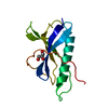
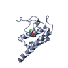
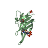
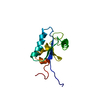
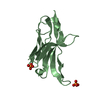
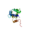
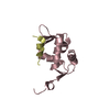
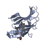
Entry Database : PDB / ID : 6g8tTitle Crystal Structures of the Single PDZ Domains from GRASP65 and their Interaction with the Golgin GM130 Golgi reassembly-stacking protein 1 Keywords / / / / / / / Function / homology Function Domain/homology Component
/ / / / / / / / / / / / / / / / / / / / / / / / / / / / / Biological species Homo sapiens (human)Method / / / Resolution : 2.67 Å Authors Jurk, C.M. / Roske, Y. / Heinemann, U. Journal : Croatica Chemica Acta / Year : 2018Title : Crystal Structures of the Single PDZ Domains from GRASP65 and their Interaction with the Golgin GM130Authors : Jurk, C.M. / Roske, Y. / Heinemann, U. History Deposition Apr 10, 2018 Deposition site / Processing site Revision 1.0 Apr 24, 2019 Provider / Type Revision 1.1 May 6, 2020 Group / Category Item _citation.country / _citation.journal_abbrev ... _citation.country / _citation.journal_abbrev / _citation.journal_id_CSD / _citation.journal_id_ISSN / _citation.pdbx_database_id_DOI / _citation.year Revision 1.2 Jan 17, 2024 Group / Database references / Refinement descriptionCategory chem_comp_atom / chem_comp_bond ... chem_comp_atom / chem_comp_bond / database_2 / pdbx_initial_refinement_model Item / _database_2.pdbx_database_accession
Show all Show less
 Movie
Movie Controller

 Yorodumi
 Open data
 Basic information
 Components
 Keywords
 Function and homology information
 Homo sapiens (human)
 X-RAY DIFFRACTION /
  Authors
 Citation
 Structure visualization
 Downloads & links
 Other downloads
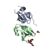


 PDBj
PDBj


 Assembly
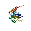


 Mass: 92.094 Da / Num. of mol.: 1 / Source method: obtained synthetically / Formula: C3H8O3
 Mass: 18.015 Da / Num. of mol.: 11 / Source method: isolated from a natural source / Formula: H2O
 Sample preparation
 / Beamline: 14.1 / Wavelength: 0.91841 Å
 Processing