 Movie
Movie Controller

[English] 日本語
 Yorodumi
- PDB-6g8c: Crystal Structure of the Amyloid-like IYQYGG segment from the R1 ... -
+ Open data

- Basic information
| Entry | Database: PDB / ID: 6g8c | ||||||
|---|---|---|---|---|---|---|---|
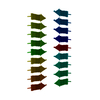
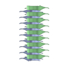
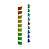
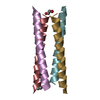
| Title | Crystal Structure of the Amyloid-like IYQYGG segment from the R1 repeat of the E. coli Biofilm-associated CsgA Curli protein | ||||||
 Components Components | Major curlin subunit | ||||||
 Keywords Keywords | PROTEIN FIBRIL / Bacterial steric-zipper cross-beta amyloid fibril from E. coli | ||||||
| Function / homology |  Function and homology information Function and homology informationAttachment of bacteria to epithelial cells / regulation of amyloid fibril formation / single-species biofilm formation / pilus / amyloid fibril formation / cell adhesion / identical protein binding Similarity search - Function | ||||||
| Biological species |  | ||||||
| Method |  X-RAY DIFFRACTION / SYNCHROTRON / MOLECULAR REPLACEMENT / molecular replacement / Resolution: 1.65 Å X-RAY DIFFRACTION / SYNCHROTRON / MOLECULAR REPLACEMENT / molecular replacement / Resolution: 1.65 Å | ||||||
| Model details | Curli | ||||||
 Authors Authors | Landau, M. / Perov, S. | ||||||
 Citation Citation | Journal: Plos Pathog. / Year: 2019 Title: Structural Insights into Curli CsgA Cross-beta Fibril Architecture Inspire Repurposing of Anti-amyloid Compounds as Anti-biofilm Agents. Authors: Perov, S. / Lidor, O. / Salinas, N. / Golan, N. / Tayeb-Fligelman, E. / Deshmukh, M. / Willbold, D. / Landau, M. | ||||||
| History |
|
- Structure visualization
| Structure viewer | Molecule: MolmilJmol/JSmol |
|---|
- Downloads & links
-Download
| PDBx/mmCIF format | 6g8c.cif.gz | 10.3 KB | Display | PDBx/mmCIF format |
|---|---|---|---|---|
| PDB format | pdb6g8c.ent.gz | 4.8 KB | Display | PDB format |
| PDBx/mmJSON format | 6g8c.json.gz | Tree view | PDBx/mmJSON format | |
| Others |  Other downloads Other downloads |
-Validation report
| Arichive directory | https://data.pdbj.org/pub/pdb/validation_reports/g8/6g8cftp://data.pdbj.org/pub/pdb/validation_reports/g8/6g8c | HTTPS FTP |
|---|
-Related structure data


-Links
 PDBj
PDBj- Assembly
| Deposited unit | 
| ||||||||
|---|---|---|---|---|---|---|---|---|---|
| 1 | 
| ||||||||
| Unit cell |
|
-Components
| #1: Protein/peptide | Mass: 699.753 Da / Num. of mol.: 1 Fragment: Amyloid spine segment IYQYGG from CsgA (residues 47-52) secreted by E. coli Source method: obtained synthetically / Details: IYQYGG from CsgA, synthesized / Source: (synth.) |
|---|---|
| #2: Water | ChemComp-HOH /  Mass: 18.015 Da / Num. of mol.: 2 / Source method: isolated from a natural source / Formula: H2O Mass: 18.015 Da / Num. of mol.: 2 / Source method: isolated from a natural source / Formula: H2O |
-Experimental details
-Experiment
| Experiment | Method: X-RAY DIFFRACTION / Number of used crystals: 1 |
|---|
- Sample preparation
| Crystal | Description: needle-like |
|---|---|
| Crystal grow | Temperature: 293 K / Method: vapor diffusion, hanging drop Details: Reservoir contained 0.1 M Sodium acetate pH 4.6 and 2.0 M Sodium formate |
-Data collection
| Diffraction | Mean temperature: 100 K | ||||||||||||||||||||||||||||||||||||||||||||||||
|---|---|---|---|---|---|---|---|---|---|---|---|---|---|---|---|---|---|---|---|---|---|---|---|---|---|---|---|---|---|---|---|---|---|---|---|---|---|---|---|---|---|---|---|---|---|---|---|---|---|
| Diffraction source | Source: SYNCHROTRON / Site: PETRA III, EMBL c/o DESY  / Beamline: P14 (MX2) / Wavelength: 0.9763 Å / Beamline: P14 (MX2) / Wavelength: 0.9763 Å | ||||||||||||||||||||||||||||||||||||||||||||||||
| Detector | Type: DECTRIS PILATUS 6M-F / Detector: PIXEL / Date: May 2, 2016 | ||||||||||||||||||||||||||||||||||||||||||||||||
| Radiation | Protocol: SINGLE WAVELENGTH / Monochromatic (M) / Laue (L): M / Scattering type: x-ray | ||||||||||||||||||||||||||||||||||||||||||||||||
| Radiation wavelength | Wavelength: 0.9763 Å / Relative weight: 1 | ||||||||||||||||||||||||||||||||||||||||||||||||
| Reflection | Resolution: 1.65→19.14 Å / Num. obs: 545 / % possible obs: 97.3 % / Redundancy: 5.231 % / Biso Wilson estimate: 19.055 Å2 / CC1/2: 0.996 / Rmerge(I) obs: 0.099 / Rrim(I) all: 0.111 / Χ2: 1.116 / Net I/σ(I): 8.66 | ||||||||||||||||||||||||||||||||||||||||||||||||
| Reflection shell | Diffraction-ID: 1
|
-Phasing
| Phasing | Method: molecular replacement | |||||||||
|---|---|---|---|---|---|---|---|---|---|---|
| Phasing MR | Model details: Phaser MODE: MR_AUTO
|
- Processing
| Software |
| |||||||||||||||||||||||||||||||||||||||||||||||||||||||
|---|---|---|---|---|---|---|---|---|---|---|---|---|---|---|---|---|---|---|---|---|---|---|---|---|---|---|---|---|---|---|---|---|---|---|---|---|---|---|---|---|---|---|---|---|---|---|---|---|---|---|---|---|---|---|---|---|
| Refinement | Method to determine structure: MOLECULAR REPLACEMENT Starting model: Ideal beta-strand Resolution: 1.65→19.14 Å / Cor.coef. Fo:Fc: 0.982 / Cor.coef. Fo:Fc free: 0.97 / SU B: 1.799 / SU ML: 0.056 / SU R Cruickshank DPI: 0.1077 / Cross valid method: THROUGHOUT / σ(F): 0 / ESU R: 0.108 / ESU R Free: 0.1 Details: HYDROGENS HAVE BEEN ADDED IN THE RIDING POSITIONS U VALUES : REFINED INDIVIDUALLY
| |||||||||||||||||||||||||||||||||||||||||||||||||||||||
| Solvent computation | Ion probe radii: 0.8 Å / Shrinkage radii: 0.8 Å / VDW probe radii: 1.2 Å | |||||||||||||||||||||||||||||||||||||||||||||||||||||||
| Displacement parameters | Biso max: 26.07 Å2 / Biso mean: 11.969 Å2 / Biso min: 8.04 Å2
| |||||||||||||||||||||||||||||||||||||||||||||||||||||||
| Refinement step | Cycle: final / Resolution: 1.65→19.14 Å
| |||||||||||||||||||||||||||||||||||||||||||||||||||||||
| Refine LS restraints |
| |||||||||||||||||||||||||||||||||||||||||||||||||||||||
| LS refinement shell | Resolution: 1.652→1.694 Å / Rfactor Rfree error: 0 / Total num. of bins used: 20
|
