 Movie
Movie Controller

[English] 日本語
 Yorodumi
- PDB-6fy3: Crystal structure of a V2-directed, RV144 vaccine-like antibody f... -
+ Open data

- Basic information
| Entry | Database: PDB / ID: 6fy3 | ||||||||||||
|---|---|---|---|---|---|---|---|---|---|---|---|---|---|
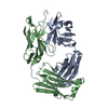
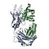
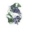
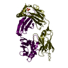
| Title | Crystal structure of a V2-directed, RV144 vaccine-like antibody from HIV-1 infection, CAP228-3D, bound to a heterologous V2 peptide | ||||||||||||
 Components Components |
| ||||||||||||
 Keywords Keywords | IMMUNE SYSTEM / Fab / HIV-1 Envelope V1V2 | ||||||||||||
| Function / homology |  Function and homology information Function and homology information: / positive regulation of establishment of T cell polarity / positive regulation of plasma membrane raft polarization / positive regulation of receptor clustering / host cell endosome membrane / clathrin-dependent endocytosis of virus by host cell / viral protein processing / fusion of virus membrane with host plasma membrane / fusion of virus membrane with host endosome membrane / apoptotic process ...: / positive regulation of establishment of T cell polarity / positive regulation of plasma membrane raft polarization / positive regulation of receptor clustering / host cell endosome membrane / clathrin-dependent endocytosis of virus by host cell / viral protein processing / fusion of virus membrane with host plasma membrane / fusion of virus membrane with host endosome membrane / apoptotic process / viral envelope / virion attachment to host cell / host cell plasma membrane / virion membrane / structural molecule activity / plasma membrane Similarity search - Function | ||||||||||||
| Biological species |  Homo sapiens (human) Homo sapiens (human)  Human immunodeficiency virus 1 Human immunodeficiency virus 1 | ||||||||||||
| Method |  X-RAY DIFFRACTION / SYNCHROTRON / MOLECULAR REPLACEMENT / Resolution: 2.6 Å X-RAY DIFFRACTION / SYNCHROTRON / MOLECULAR REPLACEMENT / Resolution: 2.6 Å | ||||||||||||
 Authors Authors | Wibmer, C.K. / Moore, P.L. / Morris, L. | ||||||||||||
| Funding support |  United States, United States,  South Africa, 3items South Africa, 3items
| ||||||||||||
 Citation Citation | Journal: Nat Commun / Year: 2018 Title: Common helical V1V2 conformations of HIV-1 Envelope expose the alpha 4 beta 7 binding site on intact virions. Authors: Wibmer, C.K. / Richardson, S.I. / Yolitz, J. / Cicala, C. / Arthos, J. / Moore, P.L. / Morris, L. | ||||||||||||
| History |
|
- Structure visualization
| Structure viewer | Molecule: MolmilJmol/JSmol |
|---|
- Downloads & links
-Download
| PDBx/mmCIF format | 6fy3.cif.gz | 484.2 KB | Display | PDBx/mmCIF format |
|---|---|---|---|---|
| PDB format | pdb6fy3.ent.gz | 405.3 KB | Display | PDB format |
| PDBx/mmJSON format | 6fy3.json.gz | Tree view | PDBx/mmJSON format | |
| Others |  Other downloads Other downloads |
-Validation report
| Arichive directory | https://data.pdbj.org/pub/pdb/validation_reports/fy/6fy3ftp://data.pdbj.org/pub/pdb/validation_reports/fy/6fy3 | HTTPS FTP |
|---|
-Related structure data
| Related structure data |  6fy1C  6fy2C  4hqqS S: Starting model for refinement C: citing same article ( |
|---|---|
| Similar structure data |


-Links
 PDBj
PDBj


- Assembly
| Deposited unit | 
| ||||||||
|---|---|---|---|---|---|---|---|---|---|
| 1 | 
| ||||||||
| 2 | 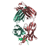
| ||||||||
| Unit cell |
|
-Components
| #1: Antibody | Mass: 25697.035 Da / Num. of mol.: 2 Source method: isolated from a genetically manipulated source Details: Donor CAP228 / Source: (gene. exp.) Homo sapiens (human) / Cell: Memory B cell / Gene: IGHV5-51 / Cell line (production host): FreeStyle 293-F / Production host: Homo sapiens (human)#2: Antibody | Mass: 23508.760 Da / Num. of mol.: 2 Source method: isolated from a genetically manipulated source Details: Donor CAP228 / Source: (gene. exp.) Homo sapiens (human) / Cell: Memory B cell / Gene: IGLV6-57 / Cell line (production host): FreeStyle 293-F / Production host: Homo sapiens (human)#3: Protein/peptide | Mass: 2344.729 Da / Num. of mol.: 2 Source method: isolated from a genetically manipulated source Source: (gene. exp.) Human immunodeficiency virus 1 / Strain: CAP45 / Gene: Env / Variant: 2.00.G3 / Production host: synthetic construct (others) / References: UniProt: C6FX86*PLUS#4: Water | ChemComp-HOH / |  Mass: 18.015 Da / Num. of mol.: 168 / Source method: isolated from a natural source / Formula: H2O Mass: 18.015 Da / Num. of mol.: 168 / Source method: isolated from a natural source / Formula: H2OHas protein modification | Y | |
|---|
-Experimental details
-Experiment
| Experiment | Method: X-RAY DIFFRACTION / Number of used crystals: 1 |
|---|
- Sample preparation
| Crystal | Density Matthews: 2.65 Å3/Da / Density % sol: 53.55 % / Description: Rods |
|---|---|
| Crystal grow | Temperature: 298.15 K / Method: vapor diffusion, hanging drop / pH: 7 Details: 0.05 M imidazole HCl (pH6.5), 0.05M HEPES NaOH (pH7.5), 20% PEG8000, 5% 2-methyl-2,4-pentanediol (MPD), 0.1 M ammonium sulphate, 5% isopropanol, supplemented up to 25% MPD as a cryoprotectant |
-Data collection
| Diffraction | Mean temperature: 100 K |
|---|---|
| Diffraction source | Source: SYNCHROTRON / Site: APS / Beamline: 22-ID / Wavelength: 1 Å |
| Detector | Type: MARMOSAIC 300 mm CCD / Detector: CCD / Date: Oct 15, 2016 |
| Radiation | Protocol: SINGLE WAVELENGTH / Monochromatic (M) / Laue (L): M / Scattering type: x-ray |
| Radiation wavelength | Wavelength: 1 Å / Relative weight: 1 |
| Reflection | Resolution: 2.6→50 Å / Num. obs: 33817 / % possible obs: 100 % / Redundancy: 6 % / Net I/σ(I): 7.5 |
| Reflection shell | Resolution: 2.6→2.64 Å / Redundancy: 3.6 % / Mean I/σ(I) obs: 3.9 / % possible all: 100 |
- Processing
| Software |
| |||||||||||||||||||||||||||||||||||||||||||||||||||||||||||||||||||||||||||||||||||||||||||
|---|---|---|---|---|---|---|---|---|---|---|---|---|---|---|---|---|---|---|---|---|---|---|---|---|---|---|---|---|---|---|---|---|---|---|---|---|---|---|---|---|---|---|---|---|---|---|---|---|---|---|---|---|---|---|---|---|---|---|---|---|---|---|---|---|---|---|---|---|---|---|---|---|---|---|---|---|---|---|---|---|---|---|---|---|---|---|---|---|---|---|---|---|
| Refinement | Method to determine structure: MOLECULAR REPLACEMENT Starting model: 4HQQ Resolution: 2.6→42.867 Å / SU ML: 0.31 / Cross valid method: FREE R-VALUE / σ(F): 1.34 / Phase error: 26.16
| |||||||||||||||||||||||||||||||||||||||||||||||||||||||||||||||||||||||||||||||||||||||||||
| Solvent computation | Shrinkage radii: 0.9 Å / VDW probe radii: 1.11 Å | |||||||||||||||||||||||||||||||||||||||||||||||||||||||||||||||||||||||||||||||||||||||||||
| Refinement step | Cycle: LAST / Resolution: 2.6→42.867 Å
| |||||||||||||||||||||||||||||||||||||||||||||||||||||||||||||||||||||||||||||||||||||||||||
| Refine LS restraints |
| |||||||||||||||||||||||||||||||||||||||||||||||||||||||||||||||||||||||||||||||||||||||||||
| LS refinement shell |
| |||||||||||||||||||||||||||||||||||||||||||||||||||||||||||||||||||||||||||||||||||||||||||
| Refinement TLS params. | Method: refined / Origin x: -34.9625 Å / Origin y: -1.214 Å / Origin z: 15.8981 Å
| |||||||||||||||||||||||||||||||||||||||||||||||||||||||||||||||||||||||||||||||||||||||||||
| Refinement TLS group | Selection details: all |
