| 登録情報 | データベース: PDB / ID: 6fuf
|
|---|
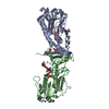
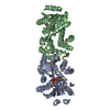
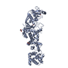
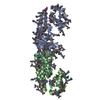
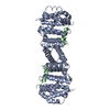
| タイトル | Crystal structure of the rhodopsin-mini-Go complex |
|---|
 要素 要素 | - Guanine nucleotide-binding protein G(o) subunit alpha
- Rhodopsin
|
|---|
 キーワード キーワード | SIGNALING PROTEIN / GPCR Complex Rhodopsin |
|---|
| 機能・相同性 |  機能・相同性情報 機能・相同性情報
Opsins / VxPx cargo-targeting to cilium / sperm head plasma membrane / rod bipolar cell differentiation / absorption of visible light / opsin binding / The canonical retinoid cycle in rods (twilight vision) / G protein-coupled opsin signaling pathway / photoreceptor inner segment membrane / podosome assembly ...Opsins / VxPx cargo-targeting to cilium / sperm head plasma membrane / rod bipolar cell differentiation / absorption of visible light / opsin binding / The canonical retinoid cycle in rods (twilight vision) / G protein-coupled opsin signaling pathway / photoreceptor inner segment membrane / podosome assembly / 11-cis retinal binding / G protein-coupled photoreceptor activity / rod photoreceptor outer segment / cellular response to light stimulus / G protein-coupled receptor complex / Inactivation, recovery and regulation of the phototransduction cascade / thermotaxis / Activation of the phototransduction cascade / mu-type opioid receptor binding / outer membrane / detection of temperature stimulus involved in thermoception / corticotropin-releasing hormone receptor 1 binding / response to light intensity / photoreceptor cell maintenance / arrestin family protein binding / vesicle docking involved in exocytosis / G protein-coupled dopamine receptor signaling pathway / photoreceptor outer segment membrane / G alpha (i) signalling events / regulation of heart contraction / parallel fiber to Purkinje cell synapse / response to light stimulus / phototransduction, visible light / G-protein alpha-subunit binding / phototransduction / photoreceptor outer segment / postsynaptic modulation of chemical synaptic transmission / adenylate cyclase regulator activity / sperm midpiece / G protein-coupled serotonin receptor binding / adenylate cyclase-inhibiting serotonin receptor signaling pathway / visual perception / muscle contraction / guanyl-nucleotide exchange factor activity / locomotory behavior / negative regulation of insulin secretion / GABA-ergic synapse / adenylate cyclase-modulating G protein-coupled receptor signaling pathway / G-protein beta/gamma-subunit complex binding / microtubule cytoskeleton organization / photoreceptor disc membrane / cell-cell junction / heterotrimeric G-protein complex / G protein activity / presynaptic membrane / cell body / Ca2+ pathway / 加水分解酵素; 酸無水物に作用; GTPに作用・細胞または細胞小器官の運動に関与 / gene expression / postsynaptic membrane / G protein-coupled receptor signaling pathway / Golgi membrane / GTPase activity / dendrite / GTP binding / glutamatergic synapse / zinc ion binding / metal ion binding / identical protein binding / membrane / plasma membrane / cytoplasm類似検索 - 分子機能 Rhodopsin, N-terminal / Amino terminal of the G-protein receptor rhodopsin / Rhodopsin / Opsin / Visual pigments (opsins) retinal binding site / Visual pigments (opsins) retinal binding site. / : / Rhopdopsin 7-helix transmembrane proteins / Rhodopsin 7-helix transmembrane proteins / G-protein alpha subunit, group I ...Rhodopsin, N-terminal / Amino terminal of the G-protein receptor rhodopsin / Rhodopsin / Opsin / Visual pigments (opsins) retinal binding site / Visual pigments (opsins) retinal binding site. / : / Rhopdopsin 7-helix transmembrane proteins / Rhodopsin 7-helix transmembrane proteins / G-protein alpha subunit, group I / Serpentine type 7TM GPCR chemoreceptor Srsx / Guanine nucleotide binding protein (G-protein), alpha subunit / G protein alpha subunit, helical insertion / G-protein alpha subunit / G-alpha domain profile. / G protein alpha subunit / G-protein coupled receptors family 1 signature. / G protein-coupled receptor, rhodopsin-like / GPCR, rhodopsin-like, 7TM / G-protein coupled receptors family 1 profile. / 7 transmembrane receptor (rhodopsin family) / P-loop containing nucleotide triphosphate hydrolases / Up-down Bundle / Rossmann fold / P-loop containing nucleoside triphosphate hydrolase / 3-Layer(aba) Sandwich / Mainly Alpha / Alpha Beta類似検索 - ドメイン・相同性 RETINAL / Rhodopsin / Guanine nucleotide-binding protein G(o) subunit alpha類似検索 - 構成要素 |
|---|
| 生物種 |   Bos taurus (ウシ) Bos taurus (ウシ)
 Homo sapiens (ヒト) Homo sapiens (ヒト) |
|---|
| 手法 |  X線回折 / シンクロトロン / 分子置換 / 解像度: 3.117 Å X線回折 / シンクロトロン / 分子置換 / 解像度: 3.117 Å |
|---|
 データ登録者 データ登録者 | Tsai, C.-J. / Weinert, T. / Muehle, J. / Pamula, F. / Nehme, R. / Flock, T. / Nogly, P. / Edwards, P.C. / Carpenter, B. / Gruhl, T. ...Tsai, C.-J. / Weinert, T. / Muehle, J. / Pamula, F. / Nehme, R. / Flock, T. / Nogly, P. / Edwards, P.C. / Carpenter, B. / Gruhl, T. / Ma, P. / Deupi, X. / Standfuss, J. / Tate, C.G. / Schertler, G.F.X. |
|---|
| 資金援助 |  スイス, スイス,  英国, 5件 英国, 5件 | 組織 | 認可番号 | 国 |
|---|
| Swiss National Science Foundation | 310030_153145 | スイス | | Swiss National Science Foundation | 310030B_173335 | スイス | | Swiss National Science Foundation | 310030B_173335 | スイス | | Swiss National Science Foundation | 31003A_159558 | スイス | | European Research Council | EMPSI, 339995 | 英国 |
|
|---|
 引用 引用 | ジャーナル: Sci Adv / 年: 2018
タイトル: Crystal structure of rhodopsin in complex with a mini-Gosheds light on the principles of G protein selectivity.
著者: Tsai, C.J. / Pamula, F. / Nehme, R. / Muhle, J. / Weinert, T. / Flock, T. / Nogly, P. / Edwards, P.C. / Carpenter, B. / Gruhl, T. / Ma, P. / Deupi, X. / Standfuss, J. / Tate, C.G. / Schertler, G.F.X. |
|---|
| 履歴 | | 登録 | 2018年2月27日 | 登録サイト: PDBE / 処理サイト: PDBE |
|---|
| 改定 1.0 | 2018年10月3日 | Provider: repository / タイプ: Initial release |
|---|
| 改定 1.1 | 2018年10月10日 | Group: Data collection / Database references / カテゴリ: citation / citation_author
Item: _citation.journal_volume / _citation.page_first ..._citation.journal_volume / _citation.page_first / _citation.page_last / _citation.pdbx_database_id_PubMed / _citation.title / _citation_author.identifier_ORCID / _citation_author.name |
|---|
| 改定 1.2 | 2020年7月29日 | Group: Data collection / Derived calculations / Structure summary
カテゴリ: chem_comp / diffrn_radiation_wavelength ...chem_comp / diffrn_radiation_wavelength / entity / pdbx_chem_comp_identifier / pdbx_entity_nonpoly / struct_conn / struct_site / struct_site_gen
Item: _chem_comp.name / _chem_comp.type ..._chem_comp.name / _chem_comp.type / _diffrn_radiation_wavelength.wavelength / _entity.pdbx_description / _pdbx_entity_nonpoly.name / _struct_conn.pdbx_role
解説: Carbohydrate remediation / Provider: repository / タイプ: Remediation |
|---|
| 改定 1.3 | 2024年1月17日 | Group: Data collection / Database references ...Data collection / Database references / Refinement description / Structure summary
カテゴリ: chem_comp / chem_comp_atom ...chem_comp / chem_comp_atom / chem_comp_bond / database_2 / pdbx_initial_refinement_model
Item: _chem_comp.pdbx_synonyms / _database_2.pdbx_DOI / _database_2.pdbx_database_accession |
|---|
| 改定 1.4 | 2024年11月13日 | Group: Structure summary
カテゴリ: pdbx_entry_details / pdbx_modification_feature |
|---|
|
|---|
 ムービー
ムービー コントローラー


 データを開く
 基本情報
基本情報 構造の表示
構造の表示 ダウンロードとリンク
 その他のダウンロード
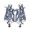


 PDBj
PDBj


 集合体
集合体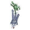


 分子量: 284.436 Da / 分子数: 1 / 由来タイプ: 合成 / 式: C20H28O / タイプ: SUBJECT OF INVESTIGATION
 タイプ: D-saccharide, beta linking / 分子量: 221.208 Da / 分子数: 1 / 由来タイプ: 組換発現 / 式: C8H15NO6
 分子量: 18.015 Da / 分子数: 1 / 由来タイプ: 天然 / 式: H2O
 試料調製
試料調製 解析
解析