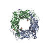
 Movie
Movie Controller


+ Open data

- Basic information
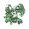
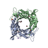
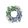
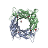
| Entry | Database: PDB / ID: 6fpn | |||||||||
|---|---|---|---|---|---|---|---|---|---|---|
| Title | Lytic transglycosylase in action | |||||||||
 Components Components | Putative soluble lytic murein transglycosylase | |||||||||
 Keywords Keywords | HYDROLASE / lytic transglycosylases / acid/base catalysis / peptidoglycan / bacteria | |||||||||
| Function / homology |  Function and homology information Function and homology informationcatalytic activity / hydrolase activity, hydrolyzing O-glycosyl compounds / periplasmic space / metal ion binding Similarity search - Function | |||||||||
| Biological species |  Neisseria meningitidis (bacteria) Neisseria meningitidis (bacteria) | |||||||||
| Method |  X-RAY DIFFRACTION / SYNCHROTRON / MOLECULAR REPLACEMENT / Resolution: 1.44 Å X-RAY DIFFRACTION / SYNCHROTRON / MOLECULAR REPLACEMENT / Resolution: 1.44 Å | |||||||||
 Authors Authors | Williams, A.H. / Hoauz, A. / Boneca, I.G. | |||||||||
 Citation Citation | Journal: J. Biol. Chem. / Year: 2018 Title: A step-by-stepin crystalloguide to bond cleavage and 1,6-anhydro-sugar product synthesis by a peptidoglycan-degrading lytic transglycosylase. Authors: Williams, A.H. / Wheeler, R. / Rateau, L. / Malosse, C. / Chamot-Rooke, J. / Haouz, A. / Taha, M.K. / Boneca, I.G. | |||||||||
| History |
|
- Structure visualization
| Structure viewer | Molecule: MolmilJmol/JSmol |
|---|
- Downloads & links
-Download
| PDBx/mmCIF format | 6fpn.cif.gz | 146.4 KB | Display | PDBx/mmCIF format |
|---|---|---|---|---|
| PDB format | pdb6fpn.ent.gz | 109.6 KB | Display | PDB format |
| PDBx/mmJSON format | 6fpn.json.gz | Tree view | PDBx/mmJSON format | |
| Others |  Other downloads Other downloads |
-Validation report
| Summary document | 6fpn_validation.pdf.gz | 412.9 KB | Display | wwPDB validaton report |
|---|---|---|---|---|
| Full document | 6fpn_full_validation.pdf.gz | 418.8 KB | Display | |
| Data in XML | 6fpn_validation.xml.gz | 13.7 KB | Display | |
| Data in CIF | 6fpn_validation.cif.gz | 24.9 KB | Display | |
| Arichive directory | https://data.pdbj.org/pub/pdb/validation_reports/fp/6fpnftp://data.pdbj.org/pub/pdb/validation_reports/fp/6fpn | HTTPS FTP |
-Related structure data
| Related structure data | 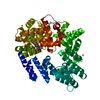 5o1jC  5o24C  5o29C 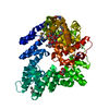 5o2nC  502nS C: citing same article ( S: Starting model for refinement |
|---|---|
| Similar structure data |


-Links
 PDBj
PDBj


- Assembly
| Deposited unit | 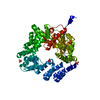
| ||||||||
|---|---|---|---|---|---|---|---|---|---|
| 1 |
| ||||||||
| Unit cell |
|
-Components
| #1: Protein | Mass: 64768.680 Da / Num. of mol.: 1 Source method: isolated from a genetically manipulated source Source: (gene. exp.) Neisseria meningitidis (bacteria) / Strain: MC58 / Gene: NMB1949 / Plasmid: PGEX-4T1 / Production host: | ||
|---|---|---|---|
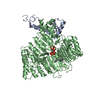
| #2: Chemical | ChemComp-SO4 /   Mass: 96.063 Da / Num. of mol.: 7 / Source method: obtained synthetically / Formula: SO4 Mass: 96.063 Da / Num. of mol.: 7 / Source method: obtained synthetically / Formula: SO4#3: Water | ChemComp-HOH / |  Mass: 18.015 Da / Num. of mol.: 721 / Source method: isolated from a natural source / Formula: H2O Mass: 18.015 Da / Num. of mol.: 721 / Source method: isolated from a natural source / Formula: H2O |
-Experimental details
-Experiment
| Experiment | Method: X-RAY DIFFRACTION / Number of used crystals: 1 |
|---|
- Sample preparation
| Crystal | Density Matthews: 2.35 Å3/Da / Density % sol: 47.73 % |
|---|---|
| Crystal grow | Temperature: 291 K / Method: vapor diffusion, hanging drop / pH: 7.5 / Details: 0.1 M 2% (v/v) PEG 400 0.1 M Hepes pH. 7.5 |
-Data collection
| Diffraction | Mean temperature: 100 K |
|---|---|
| Diffraction source | Source: SYNCHROTRON / Site: SOLEIL  / Beamline: PROXIMA 1 / Wavelength: 1 Å / Beamline: PROXIMA 1 / Wavelength: 1 Å |
| Detector | Type: DECTRIS PILATUS 6M / Detector: PIXEL / Date: Nov 15, 2012 |
| Radiation | Protocol: SINGLE WAVELENGTH / Monochromatic (M) / Laue (L): M / Scattering type: x-ray |
| Radiation wavelength | Wavelength: 1 Å / Relative weight: 1 |
| Reflection | Resolution: 1.4→62.96 Å / Num. obs: 117919 / % possible obs: 97.42 % / Redundancy: 4.7 % / Rmerge(I) obs: 0.063 / Net I/σ(I): 12.16 |
| Reflection shell | Resolution: 1.4→1.449 Å |
- Processing
| Software |
| ||||||||||||
|---|---|---|---|---|---|---|---|---|---|---|---|---|---|
| Refinement | Method to determine structure: MOLECULAR REPLACEMENT Starting model: 502N Resolution: 1.44→62.96 Å / Cross valid method: FREE R-VALUE /
| ||||||||||||
| Refinement step | Cycle: LAST / Resolution: 1.44→62.96 Å
|
