 Movie
Movie Controller


+ Open data

- Basic information
| Entry | Database: PDB / ID: 6fnl | ||||||
|---|---|---|---|---|---|---|---|
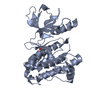
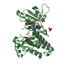
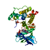
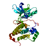
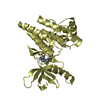
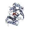
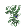
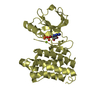
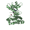
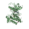
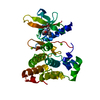
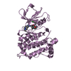
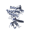
| Title | Crystal Structure of Ephrin B4 (EphB4) Receptor Protein Kinase | ||||||
 Components Components | Ephrin type-B receptor 4 | ||||||
 Keywords Keywords | TRANSFERASE / Protein Tyrosine Kinase | ||||||
| Function / homology |  Function and homology information Function and homology informationephrin receptor activity / cell migration involved in sprouting angiogenesis / EPH-Ephrin signaling / Ephrin signaling / heart morphogenesis / EPH-ephrin mediated repulsion of cells / ephrin receptor signaling pathway / EPHB-mediated forward signaling / transmembrane receptor protein tyrosine kinase activity / receptor protein-tyrosine kinase ...ephrin receptor activity / cell migration involved in sprouting angiogenesis / EPH-Ephrin signaling / Ephrin signaling / heart morphogenesis / EPH-ephrin mediated repulsion of cells / ephrin receptor signaling pathway / EPHB-mediated forward signaling / transmembrane receptor protein tyrosine kinase activity / receptor protein-tyrosine kinase / angiogenesis / signaling receptor complex / cell adhesion / extracellular exosome / extracellular region / ATP binding / plasma membrane / cytosol Similarity search - Function | ||||||
| Biological species |  Homo sapiens (human) Homo sapiens (human) | ||||||
| Method |  X-RAY DIFFRACTION / SYNCHROTRON / MOLECULAR REPLACEMENT / Resolution: 1.269 Å X-RAY DIFFRACTION / SYNCHROTRON / MOLECULAR REPLACEMENT / Resolution: 1.269 Å | ||||||
 Authors Authors | Kudlinzki, D. / Troester, A. / Witt, K. / Linhard, V.L. / Saxena, K. / Schwalbe, H. | ||||||
| Funding support |  Germany, 1items Germany, 1items
| ||||||
 Citation Citation | Journal: ChemMedChem / Year: 2018 Title: NVP-BHG712: Effects of Regioisomers on the Affinity and Selectivity toward the EPHrin Family. Authors: Troster, A. / Heinzlmeir, S. / Berger, B.T. / Gande, S.L. / Saxena, K. / Sreeramulu, S. / Linhard, V. / Nasiri, A.H. / Bolte, M. / Muller, S. / Kuster, B. / Medard, G. / Kudlinzki, D. / Schwalbe, H. | ||||||
| History |
|
- Structure visualization
| Structure viewer | Molecule: MolmilJmol/JSmol |
|---|
- Downloads & links
-Download
| PDBx/mmCIF format | 6fnl.cif.gz | 74.4 KB | Display | PDBx/mmCIF format |
|---|---|---|---|---|
| PDB format | pdb6fnl.ent.gz | 53.6 KB | Display | PDB format |
| PDBx/mmJSON format | 6fnl.json.gz | Tree view | PDBx/mmJSON format | |
| Others |  Other downloads Other downloads |
-Validation report
| Arichive directory | https://data.pdbj.org/pub/pdb/validation_reports/fn/6fnlftp://data.pdbj.org/pub/pdb/validation_reports/fn/6fnl | HTTPS FTP |
|---|
-Related structure data
| Related structure data | 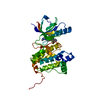 6fnfC 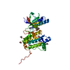 6fngC 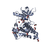 6fnhC 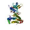 6fniC 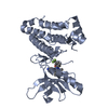 6fnjC  6fnkC 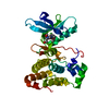 6fnmC 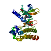 2vwuS S: Starting model for refinement C: citing same article ( |
|---|---|
| Similar structure data |


-Links
 PDBj
PDBj


- Assembly
| Deposited unit | 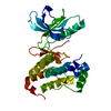
| ||||||||
|---|---|---|---|---|---|---|---|---|---|
| 1 |
| ||||||||
| Unit cell |
|
-Components
| #1: Protein | Mass: 33488.359 Da / Num. of mol.: 1 Source method: isolated from a genetically manipulated source Source: (gene. exp.) Homo sapiens (human) / Gene: EPHB4, HTK, MYK1, TYRO11 / Production host:  References: UniProt: P54760, receptor protein-tyrosine kinase |
|---|---|
| #2: Water | ChemComp-HOH /  Mass: 18.015 Da / Num. of mol.: 265 / Source method: isolated from a natural source / Formula: H2O Mass: 18.015 Da / Num. of mol.: 265 / Source method: isolated from a natural source / Formula: H2O |
-Experimental details
-Experiment
| Experiment | Method: X-RAY DIFFRACTION / Number of used crystals: 1 |
|---|
- Sample preparation
| Crystal | Density Matthews: 2.1 Å3/Da / Density % sol: 41.5 % |
|---|---|
| Crystal grow | Temperature: 291 K / Method: vapor diffusion, sitting drop / pH: 7.5 / Details: 25 % PEG5000 MME, 15 % Glycerol, 0.1 M TRIS pH7.5 |
-Data collection
| Diffraction | Mean temperature: 100 K |
|---|---|
| Diffraction source | Source: SYNCHROTRON / Site: BESSY / Beamline: 14.1 / Wavelength: 0.9184 Å |
| Detector | Type: DECTRIS PILATUS 6M / Detector: PIXEL / Date: Jan 17, 2018 |
| Radiation | Protocol: SINGLE WAVELENGTH / Monochromatic (M) / Laue (L): M / Scattering type: x-ray |
| Radiation wavelength | Wavelength: 0.9184 Å / Relative weight: 1 |
| Reflection | Resolution: 1.269→43.112 Å / Num. obs: 74570 / % possible obs: 97.8 % / Redundancy: 6.69 % / Biso Wilson estimate: 28.32 Å2 / CC1/2: 0.999 / Rrim(I) all: 0.069 / Net I/σ(I): 12.14 |
| Reflection shell | Resolution: 1.269→1.34 Å / Redundancy: 6.4 % / Mean I/σ(I) obs: 0.23 / Num. unique obs: 11399 / CC1/2: 0.126 / % possible all: 93.1 |
- Processing
| Software |
| ||||||||||||||||||||||||||||||||||||||||||||||||||||||||||||||||||||||||||||||||||||||||||||||||||||||||||||||||
|---|---|---|---|---|---|---|---|---|---|---|---|---|---|---|---|---|---|---|---|---|---|---|---|---|---|---|---|---|---|---|---|---|---|---|---|---|---|---|---|---|---|---|---|---|---|---|---|---|---|---|---|---|---|---|---|---|---|---|---|---|---|---|---|---|---|---|---|---|---|---|---|---|---|---|---|---|---|---|---|---|---|---|---|---|---|---|---|---|---|---|---|---|---|---|---|---|---|---|---|---|---|---|---|---|---|---|---|---|---|---|---|---|---|
| Refinement | Method to determine structure: MOLECULAR REPLACEMENT Starting model: 2VWU Resolution: 1.269→43.112 Å / SU ML: 0.31 / Cross valid method: FREE R-VALUE / σ(F): 1.33 / Phase error: 31.7
| ||||||||||||||||||||||||||||||||||||||||||||||||||||||||||||||||||||||||||||||||||||||||||||||||||||||||||||||||
| Solvent computation | Shrinkage radii: 0.9 Å / VDW probe radii: 1.11 Å | ||||||||||||||||||||||||||||||||||||||||||||||||||||||||||||||||||||||||||||||||||||||||||||||||||||||||||||||||
| Refinement step | Cycle: LAST / Resolution: 1.269→43.112 Å
| ||||||||||||||||||||||||||||||||||||||||||||||||||||||||||||||||||||||||||||||||||||||||||||||||||||||||||||||||
| Refine LS restraints |
| ||||||||||||||||||||||||||||||||||||||||||||||||||||||||||||||||||||||||||||||||||||||||||||||||||||||||||||||||
| LS refinement shell |
|
