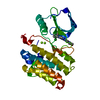
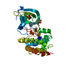
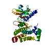
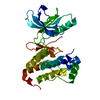
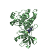
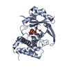
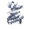
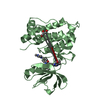
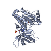
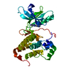
[English] 日本語
Yorodumi
- PDB-4bb4: ephB4 kinase domain inhibitor complex -

+
Open data


ID or keywords:

Loading...

-
Basic information

Entry
Database: PDB / ID: 4bb4
TitleephB4 kinase domain inhibitor complex
ComponentsEPHRIN TYPE-B RECEPTOR 4
KeywordsTRANSFERASE / UNPHOSPHORYLATED
Function / homology
Function and homology information


ephrin receptor activity / cell migration involved in sprouting angiogenesis / EPH-Ephrin signaling / Ephrin signaling / EPH-ephrin mediated repulsion of cells / ephrin receptor signaling pathway / heart morphogenesis / EPHB-mediated forward signaling / transmembrane receptor protein tyrosine kinase activity / receptor protein-tyrosine kinase ...ephrin receptor activity / cell migration involved in sprouting angiogenesis / EPH-Ephrin signaling / Ephrin signaling / EPH-ephrin mediated repulsion of cells / ephrin receptor signaling pathway / heart morphogenesis / EPHB-mediated forward signaling / transmembrane receptor protein tyrosine kinase activity / receptor protein-tyrosine kinase / angiogenesis / protein autophosphorylation / receptor complex / cell adhesion / extracellular exosome / extracellular region / ATP binding / plasma membrane / cytosol
Similarity search - Function
Ephrin type-B receptor 4, ligand binding domain / EPH-B4, SAM domain / Tyrosine-protein kinase ephrin type A/B receptor-like / Tyrosine-protein kinase ephrin type A/B receptor-like / Ephrin receptor type-A /type-B / Ephrin receptor ligand binding domain / Tyrosine-protein kinase, receptor class V, conserved site / Ephrin receptor, transmembrane domain / Ephrin receptor ligand binding domain / Ephrin type-A receptor 2 transmembrane domain ...Ephrin type-B receptor 4, ligand binding domain / EPH-B4, SAM domain / Tyrosine-protein kinase ephrin type A/B receptor-like / Tyrosine-protein kinase ephrin type A/B receptor-like / Ephrin receptor type-A /type-B / Ephrin receptor ligand binding domain / Tyrosine-protein kinase, receptor class V, conserved site / Ephrin receptor, transmembrane domain / Ephrin receptor ligand binding domain / Ephrin type-A receptor 2 transmembrane domain / Receptor tyrosine kinase class V signature 1. / Receptor tyrosine kinase class V signature 2. / Eph receptor ligand-binding domain profile. / Ephrin receptor ligand binding domain / Putative ephrin-receptor like / SAM domain (Sterile alpha motif) / SAM domain profile. / Sterile alpha motif. / Sterile alpha motif domain / Sterile alpha motif/pointed domain superfamily / Growth factor receptor cysteine-rich domain superfamily / EGF-like domain signature 2. / Fibronectin type III domain / Fibronectin type 3 domain / Fibronectin type-III domain profile. / Galactose-binding-like domain superfamily / Fibronectin type III / Fibronectin type III superfamily / Tyrosine-protein kinase, catalytic domain / Tyrosine kinase, catalytic domain / Tyrosine protein kinases specific active-site signature. / Tyrosine-protein kinase, active site / Protein tyrosine and serine/threonine kinase / Serine-threonine/tyrosine-protein kinase, catalytic domain / Transferase(Phosphotransferase) domain 1 / Transferase(Phosphotransferase); domain 1 / Phosphorylase Kinase; domain 1 / Phosphorylase Kinase; domain 1 / Protein kinase, ATP binding site / Protein kinases ATP-binding region signature. / Immunoglobulin-like fold / Protein kinase domain profile. / Protein kinase domain / Protein kinase-like domain superfamily / 2-Layer Sandwich / Orthogonal Bundle / Mainly Alpha / Alpha Beta
Similarity search - Domain/homology
Chem-32W / Ephrin type-B receptor 4
Similarity search - Component
Biological speciesHOMO SAPIENS (human)
MethodX-RAY DIFFRACTION / MOLECULAR REPLACEMENT / Resolution: 1.65 Å
AuthorsRead, J. / Brassington, C.A. / Green, I. / McCall, E.J. / Valentine, A.L.
CitationJournal: J.Med.Chem. / Year: 2013
Title: Discovery and Optimization of a Novel Series of Potent Mutant B-Raf V600E Selective Kinase Inhibitors.
Authors: Vasbinder, M.M. / Aquila, B. / Augustin, M. / Chen, H. / Cheung, T. / Cook, D. / Drew, L. / Fauber, B.P. / Glossop, S. / Grondine, M. / Hennessy, E.J. / Johannes, J. / Lee, S. / Lyne, P.D. / ...Authors: Vasbinder, M.M. / Aquila, B. / Augustin, M. / Chen, H. / Cheung, T. / Cook, D. / Drew, L. / Fauber, B.P. / Glossop, S. / Grondine, M. / Hennessy, E.J. / Johannes, J. / Lee, S. / Lyne, P.D. / Mortl, M. / Omer, C. / Palakurthi, S. / Pontz, T. / Read, J. / Sha, L. / Shen, M. / Steinbacher, S. / Wang, H. / Wu, A. / Ye, M.
History
DepositionSep 19, 2012Deposition site: PDBE / Processing site: PDBE
Revision 1.0Feb 27, 2013Provider: repository / Type: Initial release
Revision 1.1Apr 10, 2013Group: Database references / Derived calculations
Revision 1.2Apr 4, 2018Group: Data collection / Category: diffrn_source / Item: _diffrn_source.type
Revision 1.3Apr 24, 2019Group: Data collection / Source and taxonomy / Category: entity_src_gen / Item: _entity_src_gen.pdbx_host_org_cell_line

-
Structure visualization

Structure viewerMolecule:
MolmilJmol/JSmol

Downloads & links

-
Assembly

Deposited unit
A: EPHRIN TYPE-B RECEPTOR 4
hetero molecules


Theoretical massNumber of molelcules
Total (without water)34,3834
Polymers33,9351
Non-polymers4483
Water4,216234
1


  • Idetical with deposited unit
  • defined by author&software
TypeNameSymmetry operationNumber
identity operation1_555x,y,z1
MethodPISA
Unit cell
Length a, b, c (Å)46.421, 53.329, 61.209
Angle α, β, γ (deg.)90.00, 111.50, 90.00
Int Tables number4
Space group name H-MP1211

-
Components

#1: Protein EPHRIN TYPE-B RECEPTOR 4 / EPHB4 RECEPTOR TYROSINE KINASE / HEPATOMA TRANSMEMBRANE KINASE / TYROSINE-PROTEIN KINASE TYRO11


Mass: 33934.816 Da / Num. of mol.: 1 / Fragment: KINASE DOMAIN, RESIDUES 598-899 / Mutation: YES
Source method: isolated from a genetically manipulated source
Source: (gene. exp.) HOMO SAPIENS (human) / Plasmid: PFASTBAC / Cell line (production host): Sf21 / Production host: 
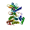
SPODOPTERA FRUGIPERDA (fall armyworm)
References: UniProt: P54760, receptor protein-tyrosine kinase
#2: Chemical ChemComp-32W / N-(2-methoxyethyl)-4-[(6-pyridin-4-ylquinazolin-2-yl)amino]benzamide


Mass: 399.445 Da / Num. of mol.: 1 / Source method: obtained synthetically / Formula: C23H21N5O2
#3: Chemical ChemComp-MG / MAGNESIUM ION


Mass: 24.305 Da / Num. of mol.: 2 / Source method: obtained synthetically / Formula: Mg
#4: Water ChemComp-HOH / water / Water


Mass: 18.015 Da / Num. of mol.: 234 / Source method: isolated from a natural source / Formula: H2O
Sequence detailsY774E MUTATION NOT SEEN IN ELECTRON DENSITY

-
Experimental details

-
Experiment

ExperimentMethod: X-RAY DIFFRACTION / Number of used crystals: 1

-
Sample preparation

CrystalDensity Matthews: 2.08 Å3/Da / Density % sol: 40.79 % / Description: NONE
Crystal growpH: 7.4 / Details: pH 7.4

-
Data collection

DiffractionMean temperature: 100 K
Diffraction sourceSource: ROTATING ANODE / Type: RIGAKU FR-E / Wavelength: 1.5418
DetectorType: RIGAKU CCD / Detector: CCD / Date: May 2, 2006 / Details: MIRRORS
RadiationMonochromator: NI FILTER / Protocol: SINGLE WAVELENGTH / Monochromatic (M) / Laue (L): M / Scattering type: x-ray
Radiation wavelengthWavelength: 1.5418 Å / Relative weight: 1
ReflectionResolution: 1.65→100 Å / Num. obs: 29124 / % possible obs: 86.7 % / Observed criterion σ(I): 2 / Redundancy: 3.27 % / Biso Wilson estimate: 30.89 Å2 / Rmerge(I) obs: 0.04 / Net I/σ(I): 13.2
Reflection shellResolution: 1.65→1.7 Å / Redundancy: 1.7 % / Rmerge(I) obs: 0.36 / Mean I/σ(I) obs: 1.8 / % possible all: 33.4

-
Processing

Software
NameVersionClassification
BUSTER-TNT2.11.2refinement
d*TREKdata reduction
d*TREKdata scaling
AMoREphasing
RefinementMethod to determine structure: MOLECULAR REPLACEMENT / Resolution: 1.65→21.28 Å / Cor.coef. Fo:Fc: 0.9481 / Cor.coef. Fo:Fc free: 0.9432 / Cross valid method: THROUGHOUT / σ(F): 2 / Details: REMARK 3
RfactorNum. reflection% reflectionSelection details
Rfree0.2361 1469 5 %RANDOM
Rwork0.2072 ---
obs0.2087 29124 86.68 %-
Displacement parametersBiso mean: 39.15 Å2
Baniso -1Baniso -2Baniso -3
1-0.7676 Å20 Å23.3102 Å2
2--1.8795 Å20 Å2
3----2.6471 Å2
Refine analyzeLuzzati coordinate error obs: 0.284 Å
Refinement stepCycle: LAST / Resolution: 1.65→21.28 Å
ProteinNucleic acidLigandSolventTotal
Num. atoms2047 0 32 234 2313
LS refinement shellResolution: 1.65→1.71 Å / Total num. of bins used: 15
RfactorNum. reflection% reflection
Rfree0.3672 63 5.81 %
Rwork0.2763 1022 -
all0.2815 1085 -
obs--33.4 %

+
About Yorodumi

-
News

-
Feb 9, 2022. New format data for meta-information of EMDB entries

New format data for meta-information of EMDB entries

  • Version 3 of the EMDB header file is now the official format.
  • The previous official version 1.9 will be removed from the archive.

Related info.:EMDB header

External links:wwPDB to switch to version 3 of the EMDB data model

-
Aug 12, 2020. Covid-19 info

Covid-19 info

URL: https://pdbj.org/emnavi/covid19.php

New page: Covid-19 featured information page in EM Navigator.

Related info.:Covid-19 info / Mar 5, 2020. Novel coronavirus structure data

+
Mar 5, 2020. Novel coronavirus structure data

Novel coronavirus structure data

Related info.:Yorodumi Speices / Aug 12, 2020. Covid-19 info

External links:COVID-19 featured content - PDBj / Molecule of the Month (242):Coronavirus Proteases

+
Jan 31, 2019. EMDB accession codes are about to change! (news from PDBe EMDB page)

EMDB accession codes are about to change! (news from PDBe EMDB page)

  • The allocation of 4 digits for EMDB accession codes will soon come to an end. Whilst these codes will remain in use, new EMDB accession codes will include an additional digit and will expand incrementally as the available range of codes is exhausted. The current 4-digit format prefixed with “EMD-” (i.e. EMD-XXXX) will advance to a 5-digit format (i.e. EMD-XXXXX), and so on. It is currently estimated that the 4-digit codes will be depleted around Spring 2019, at which point the 5-digit format will come into force.
  • The EM Navigator/Yorodumi systems omit the EMD- prefix.

Related info.:Q: What is EMD? / ID/Accession-code notation in Yorodumi/EM Navigator

External links:EMDB Accession Codes are Changing Soon! / Contact to PDBj

+
Jul 12, 2017. Major update of PDB

Major update of PDB

  • wwPDB released updated PDB data conforming to the new PDBx/mmCIF dictionary.
  • This is a major update changing the version number from 4 to 5, and with Remediation, in which all the entries are updated.
  • In this update, many items about electron microscopy experimental information are reorganized (e.g. em_software).
  • Now, EM Navigator and Yorodumi are based on the updated data.

External links:wwPDB Remediation / Enriched Model Files Conforming to OneDep Data Standards Now Available in the PDB FTP Archive

-
Yorodumi

Thousand views of thousand structures

  • Yorodumi is a browser for structure data from EMDB, PDB, SASBDB, etc.
  • This page is also the successor to EM Navigator detail page, and also detail information page/front-end page for Omokage search.
  • The word "yorodu" (or yorozu) is an old Japanese word meaning "ten thousand". "mi" (miru) is to see.

Related info.:EMDB / PDB / SASBDB / Comparison of 3 databanks / Yorodumi Search / Aug 31, 2016. New EM Navigator & Yorodumi / Yorodumi Papers / Jmol/JSmol / Function and homology information / Changes in new EM Navigator and Yorodumi

Read more