 Movie
Movie Controller

[English] 日本語
 Yorodumi
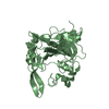
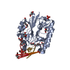
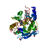
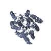
- PDB-6fk5: Structure of 3' phosphatase NExo (D146N) from Neisseria bound to ... -
+ Open data

- Basic information
| Entry | Database: PDB / ID: 6fk5 | ||||||
|---|---|---|---|---|---|---|---|
| Title | Structure of 3' phosphatase NExo (D146N) from Neisseria bound to DNA substrate in presence of magnesium ion | ||||||
 Components Components |
| ||||||
 Keywords Keywords | DNA BINDING PROTEIN / 3' phosphatase / base excision DNA repair / Mg2+ | ||||||
| Function / homology |  Function and homology information Function and homology information: / exodeoxyribonuclease III / double-stranded DNA 3'-5' DNA exonuclease activity / endonuclease activity / DNA repair / DNA damage response / DNA binding / metal ion binding / cytosol Similarity search - Function | ||||||
| Biological species |  Neisseria meningitidis MC58 (bacteria) Neisseria meningitidis MC58 (bacteria)other sequences (unknown) | ||||||
| Method |  X-RAY DIFFRACTION / SYNCHROTRON / MOLECULAR REPLACEMENT / Resolution: 2.02 Å X-RAY DIFFRACTION / SYNCHROTRON / MOLECULAR REPLACEMENT / Resolution: 2.02 Å | ||||||
 Authors Authors | Silhan, J. / Zhao, Q. / Boura, E. / Thomson, H. / Foster, A. / Tang, C.M. / Freemont, P.S. / Baldwin, G.S. | ||||||
| Funding support |  United Kingdom, 1items United Kingdom, 1items
| ||||||
 Citation Citation | Journal: Nucleic Acids Res. / Year: 2018 Title: Structural basis for recognition and repair of the 3'-phosphate by NExo, a base excision DNA repair nuclease from Neisseria meningitidis. Authors: Silhan, J. / Zhao, Q. / Boura, E. / Thomson, H. / Forster, A. / Tang, C.M. / Freemont, P.S. / Baldwin, G.S. | ||||||
| History |
|
- Structure visualization
| Structure viewer | Molecule: MolmilJmol/JSmol |
|---|
- Downloads & links
-Download
| PDBx/mmCIF format | 6fk5.cif.gz | 80.4 KB | Display | PDBx/mmCIF format |
|---|---|---|---|---|
| PDB format | pdb6fk5.ent.gz | 55.8 KB | Display | PDB format |
| PDBx/mmJSON format | 6fk5.json.gz | Tree view | PDBx/mmJSON format | |
| Others |  Other downloads Other downloads |
-Validation report
| Arichive directory | https://data.pdbj.org/pub/pdb/validation_reports/fk/6fk5ftp://data.pdbj.org/pub/pdb/validation_reports/fk/6fk5 | HTTPS FTP |
|---|
-Related structure data
| Related structure data |  6fk4C  6fkeC  2jc4S C: citing same article ( S: Starting model for refinement |
|---|---|
| Similar structure data |


-Links
 PDBj
PDBj


- Assembly
| Deposited unit | 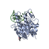
| ||||||||
|---|---|---|---|---|---|---|---|---|---|
| 1 |
| ||||||||
| Unit cell |
| ||||||||
| Components on special symmetry positions |
|
-Components
| #1: Protein | Mass: 29317.445 Da / Num. of mol.: 1 Source method: isolated from a genetically manipulated source Source: (gene. exp.) Neisseria meningitidis MC58 (bacteria) / Production host: | ||||
|---|---|---|---|---|---|
| #2: DNA chain | Mass: 5589.642 Da / Num. of mol.: 1 Source method: isolated from a genetically manipulated source Details: 3' Phosphate at the last residue / Source: (gene. exp.) other sequences (unknown) / Production host: synthetic construct (others) | ||||
| #3: Chemical |   Mass: 24.305 Da / Num. of mol.: 2 / Source method: obtained synthetically / Formula: Mg Mass: 24.305 Da / Num. of mol.: 2 / Source method: obtained synthetically / Formula: Mg#4: Chemical | ChemComp-MPD / ( |   Mass: 118.174 Da / Num. of mol.: 1 / Source method: obtained synthetically / Formula: C6H14O2 / Comment: precipitant*YM Mass: 118.174 Da / Num. of mol.: 1 / Source method: obtained synthetically / Formula: C6H14O2 / Comment: precipitant*YM#5: Water | ChemComp-HOH / |  Mass: 18.015 Da / Num. of mol.: 154 / Source method: isolated from a natural source / Formula: H2O Mass: 18.015 Da / Num. of mol.: 154 / Source method: isolated from a natural source / Formula: H2O |
-Experimental details
-Experiment
| Experiment | Method: X-RAY DIFFRACTION / Number of used crystals: 1 |
|---|
- Sample preparation
| Crystal | Density Matthews: 3.64 Å3/Da / Density % sol: 66.17 % |
|---|---|
| Crystal grow | Temperature: 277 K / Method: vapor diffusion, sitting drop / pH: 6.6 Details: 100 mM imidazole pH = 6.6, 55 mM (NH4)2SO4, 10% (w/v) PEG 8000, 16 - 19 % (v/v) MPD, 2 mM MgCl2 |
-Data collection
| Diffraction | Mean temperature: 100 K |
|---|---|
| Diffraction source | Source: SYNCHROTRON / Site: Diamond / Beamline: I24 / Wavelength: 0.9686 Å |
| Detector | Type: DECTRIS PILATUS 6M / Detector: PIXEL / Date: Feb 8, 2013 |
| Radiation | Protocol: SINGLE WAVELENGTH / Monochromatic (M) / Laue (L): M / Scattering type: x-ray |
| Radiation wavelength | Wavelength: 0.9686 Å / Relative weight: 1 |
| Reflection | Resolution: 2.02→50.43 Å / Num. obs: 35871 / % possible obs: 99 % / Redundancy: 2 % / Rmerge(I) obs: 0.0411 / Rrim(I) all: 0.05812 / Net I/σ(I): 7.4 |
| Reflection shell | Resolution: 2.02→2.08 Å / Rmerge(I) obs: 0.3141 / Rrim(I) all: 0.4442 |
- Processing
| Software |
| ||||||||||||||||||||||||||||||||||||||||||||||||||||||||||||||||||||||||||||||||||||||||||||||||||
|---|---|---|---|---|---|---|---|---|---|---|---|---|---|---|---|---|---|---|---|---|---|---|---|---|---|---|---|---|---|---|---|---|---|---|---|---|---|---|---|---|---|---|---|---|---|---|---|---|---|---|---|---|---|---|---|---|---|---|---|---|---|---|---|---|---|---|---|---|---|---|---|---|---|---|---|---|---|---|---|---|---|---|---|---|---|---|---|---|---|---|---|---|---|---|---|---|---|---|---|
| Refinement | Method to determine structure: MOLECULAR REPLACEMENT Starting model: 2JC4 Resolution: 2.02→50.43 Å / SU ML: 0.22 / Cross valid method: FREE R-VALUE / σ(F): 1.34 / Phase error: 32.06
| ||||||||||||||||||||||||||||||||||||||||||||||||||||||||||||||||||||||||||||||||||||||||||||||||||
| Solvent computation | Shrinkage radii: 0.9 Å / VDW probe radii: 1.11 Å | ||||||||||||||||||||||||||||||||||||||||||||||||||||||||||||||||||||||||||||||||||||||||||||||||||
| Refinement step | Cycle: LAST / Resolution: 2.02→50.43 Å
| ||||||||||||||||||||||||||||||||||||||||||||||||||||||||||||||||||||||||||||||||||||||||||||||||||
| Refine LS restraints |
| ||||||||||||||||||||||||||||||||||||||||||||||||||||||||||||||||||||||||||||||||||||||||||||||||||
| LS refinement shell |
|
