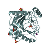
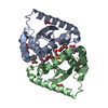
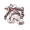
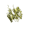
perineuronal net / regulation of oligodendrocyte progenitor proliferation / MDK and PTN in ALK signaling / transmembrane receptor protein tyrosine phosphatase activity / Other interleukin signaling / protein dephosphorylation / regulation of myelination / oligodendrocyte differentiation / peptidyl-tyrosine dephosphorylation / positive regulation of oligodendrocyte differentiation ...perineuronal net / regulation of oligodendrocyte progenitor proliferation / MDK and PTN in ALK signaling / transmembrane receptor protein tyrosine phosphatase activity / Other interleukin signaling / protein dephosphorylation / regulation of myelination / oligodendrocyte differentiation / peptidyl-tyrosine dephosphorylation / positive regulation of oligodendrocyte differentiation / hematopoietic progenitor cell differentiation / axonogenesis / protein-tyrosine-phosphatase / protein tyrosine phosphatase activity / central nervous system development / integrin binding / neuron projection development / negative regulation of neuron apoptotic process / learning or memory / synapse / signal transduction / extracellular region / plasma membrane Similarity search - Function
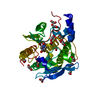
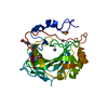
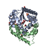
Receptor-type tyrosine-protein phosphatase, carbonic anhydrase domain / : / Protein tyrosine phosphatase, catalytic domain / PTP type protein phosphatase domain profile. / Protein-tyrosine phosphatase / Tyrosine-specific protein phosphatase, PTPase domain / Carbonic Anhydrase II / Alpha carbonic anhydrase / Protein-tyrosine phosphatase, catalytic / Protein tyrosine phosphatase, catalytic domain motif ...Receptor-type tyrosine-protein phosphatase, carbonic anhydrase domain / : / Protein tyrosine phosphatase, catalytic domain / PTP type protein phosphatase domain profile. / Protein-tyrosine phosphatase / Tyrosine-specific protein phosphatase, PTPase domain / Carbonic Anhydrase II / Alpha carbonic anhydrase / Protein-tyrosine phosphatase, catalytic / Protein tyrosine phosphatase, catalytic domain motif / Tyrosine specific protein phosphatases active site. / Protein-tyrosine phosphatase, active site / Tyrosine specific protein phosphatases domain profile. / Tyrosine-specific protein phosphatases domain / Protein-tyrosine phosphatase-like / Fibronectin type III domain / Alpha carbonic anhydrase domain / Alpha carbonic anhydrase domain superfamily / Eukaryotic-type carbonic anhydrase / Alpha-carbonic anhydrases profile. / Eukaryotic-type carbonic anhydrase / Fibronectin type 3 domain / Fibronectin type-III domain profile. / Fibronectin type III / Fibronectin type III superfamily / Roll / Immunoglobulin-like fold / Alpha Beta Similarity search - Domain/homology
In the structure databanks used in Yorodumi, some data are registered as the other names, "COVID-19 virus" and "2019-nCoV". Here are the details of the virus and the list of structure data.
Jan 31, 2019. EMDB accession codes are about to change! (news from PDBe EMDB page)
EMDB accession codes are about to change! (news from PDBe EMDB page)
The allocation of 4 digits for EMDB accession codes will soon come to an end. Whilst these codes will remain in use, new EMDB accession codes will include an additional digit and will expand incrementally as the available range of codes is exhausted. The current 4-digit format prefixed with “EMD-” (i.e. EMD-XXXX) will advance to a 5-digit format (i.e. EMD-XXXXX), and so on. It is currently estimated that the 4-digit codes will be depleted around Spring 2019, at which point the 5-digit format will come into force.
The EM Navigator/Yorodumi systems omit the EMD- prefix.
Related info.:Q: What is EMD? / ID/Accession-code notation in Yorodumi/EM Navigator
Yorodumi is a browser for structure data from EMDB, PDB, SASBDB, etc.
This page is also the successor to EM Navigator detail page, and also detail information page/front-end page for Omokage search.
The word "yorodu" (or yorozu) is an old Japanese word meaning "ten thousand". "mi" (miru) is to see.
Related info.:EMDB / PDB / SASBDB / Comparison of 3 databanks / Yorodumi Search / Aug 31, 2016. New EM Navigator & Yorodumi / Yorodumi Papers / Jmol/JSmol / Function and homology information / Changes in new EM Navigator and Yorodumi
 Movie
Movie Controller


 Open data
 Basic information
 Components
 Keywords
 Function and homology information
 Homo sapiens (human)
 X-RAY DIFFRACTION /
  Authors
 Citation
 Structure visualization
 Downloads & links
 Other downloads
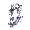


 PDBj
PDBj


 Assembly

 Mass: 18.015 Da / Num. of mol.: 222 / Source method: isolated from a natural source / Formula: H2O
 Sample preparation
 / Beamline: 22-ID / Wavelength: 1 Å
 Processing