 Movie
Movie Controller

[English] 日本語
 Yorodumi
- PDB-4zwn: Crystal Structure of a Soluble Variant of the Monoglyceride Lipas... -
+ Open data

- Basic information
| Entry | Database: PDB / ID: 4zwn | |||||||||
|---|---|---|---|---|---|---|---|---|---|---|
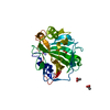
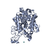
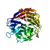
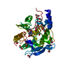
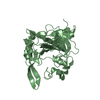
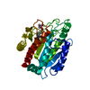
| Title | Crystal Structure of a Soluble Variant of the Monoglyceride Lipase from Saccharomyces Cerevisiae | |||||||||
 Components Components | Monoglyceride lipase | |||||||||
 Keywords Keywords | HYDROLASE / Monoglyceride Lipase / Monoacylglycerol Lipase / Serine Hydrolase / Alpha/Beta Hydrolase | |||||||||
| Function / homology |  Function and homology information Function and homology informationAcyl chain remodeling of DAG and TAG / Arachidonate production from DAG / acylglycerol lipase / monoacylglycerol lipase activity / triglyceride catabolic process / serine hydrolase activity / triglyceride metabolic process / lipid droplet / cell periphery / mitochondrial outer membrane ...Acyl chain remodeling of DAG and TAG / Arachidonate production from DAG / acylglycerol lipase / monoacylglycerol lipase activity / triglyceride catabolic process / serine hydrolase activity / triglyceride metabolic process / lipid droplet / cell periphery / mitochondrial outer membrane / endoplasmic reticulum / mitochondrion / membrane / plasma membrane / cytoplasm Similarity search - Function | |||||||||
| Biological species |  | |||||||||
| Method |  X-RAY DIFFRACTION / SYNCHROTRON / MAD / Resolution: 2.491 Å X-RAY DIFFRACTION / SYNCHROTRON / MAD / Resolution: 2.491 Å | |||||||||
 Authors Authors | Aschauer, P. / Rengachari, S. / Gruber, K. / Oberer, M. | |||||||||
| Funding support |  Austria, 2items Austria, 2items
| |||||||||
 Citation Citation | Journal: Biochim.Biophys.Acta / Year: 2016 Title: Crystal structure of the Saccharomyces cerevisiae monoglyceride lipase Yju3p. Authors: Aschauer, P. / Rengachari, S. / Lichtenegger, J. / Schittmayer, M. / Das, K.M. / Mayer, N. / Breinbauer, R. / Birner-Gruenberger, R. / Gruber, C.C. / Zimmermann, R. / Gruber, K. / Oberer, M. | |||||||||
| History |
|
- Structure visualization
| Structure viewer | Molecule: MolmilJmol/JSmol |
|---|
- Downloads & links
-Download
| PDBx/mmCIF format | 4zwn.cif.gz | 261.3 KB | Display | PDBx/mmCIF format |
|---|---|---|---|---|
| PDB format | pdb4zwn.ent.gz | 210.7 KB | Display | PDB format |
| PDBx/mmJSON format | 4zwn.json.gz | Tree view | PDBx/mmJSON format | |
| Others |  Other downloads Other downloads |
-Validation report
| Arichive directory | https://data.pdbj.org/pub/pdb/validation_reports/zw/4zwnftp://data.pdbj.org/pub/pdb/validation_reports/zw/4zwn | HTTPS FTP |
|---|
-Related structure data

-Links
 PDBj
PDBj- Assembly
| Deposited unit | 
| ||||||||
|---|---|---|---|---|---|---|---|---|---|
| 1 | 
| ||||||||
| 2 | 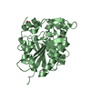
| ||||||||
| 3 | 
| ||||||||
| 4 | 
| ||||||||
| Unit cell |
|
-Components
| #1: Protein | Mass: 38040.082 Da / Num. of mol.: 4 / Fragment: UNP residues 2-313 / Mutation: Q264R, L175S Source method: isolated from a genetically manipulated source Source: (gene. exp.) Strain: ATCC 204508 / S288c / Gene: YJU3, YKL094W, YKL441 / Plasmid: pPRoEx Htb / Production host:  #2: Chemical | ChemComp-NO3 / |   Mass: 62.005 Da / Num. of mol.: 1 / Source method: obtained synthetically / Formula: NO3 Mass: 62.005 Da / Num. of mol.: 1 / Source method: obtained synthetically / Formula: NO3#3: Chemical |   Mass: 96.063 Da / Num. of mol.: 2 / Source method: obtained synthetically / Formula: SO4 Mass: 96.063 Da / Num. of mol.: 2 / Source method: obtained synthetically / Formula: SO4#4: Chemical | ChemComp-NA / |   Mass: 22.990 Da / Num. of mol.: 1 / Source method: obtained synthetically / Formula: Na Mass: 22.990 Da / Num. of mol.: 1 / Source method: obtained synthetically / Formula: Na#5: Water | ChemComp-HOH / |  Mass: 18.015 Da / Num. of mol.: 408 / Source method: isolated from a natural source / Formula: H2O Mass: 18.015 Da / Num. of mol.: 408 / Source method: isolated from a natural source / Formula: H2O |
|---|
-Experimental details
-Experiment
| Experiment | Method: X-RAY DIFFRACTION / Number of used crystals: 1 |
|---|
- Sample preparation
| Crystal | Density Matthews: 2.48 Å3/Da / Density % sol: 50.5 % |
|---|---|
| Crystal grow | Temperature: 293.15 K / Method: vapor diffusion, sitting drop / pH: 8.7 Details: 0.1 M Bicine/Trizma base pH 8.7, 10% w/v PEG 20 000, 20% v/v PEG MME 550 and 0.03 M sodium nitrate, 0.03 M disodium hydrogen phosphate, 0.03 M ammonium sulphate. Microseeding |
-Data collection
| Diffraction | Mean temperature: 100 K | |||||||||||||||||||||||||||
|---|---|---|---|---|---|---|---|---|---|---|---|---|---|---|---|---|---|---|---|---|---|---|---|---|---|---|---|---|
| Diffraction source | Source: SYNCHROTRON / Site: ESRF  / Beamline: ID29 / Wavelength: 0.9999 Å / Beamline: ID29 / Wavelength: 0.9999 Å | |||||||||||||||||||||||||||
| Detector | Type: MARMOSAIC 225 mm CCD / Detector: CCD / Date: Aug 20, 2012 | |||||||||||||||||||||||||||
| Radiation | Protocol: SINGLE WAVELENGTH / Monochromatic (M) / Laue (L): M / Scattering type: x-ray | |||||||||||||||||||||||||||
| Radiation wavelength | Wavelength: 0.9999 Å / Relative weight: 1 | |||||||||||||||||||||||||||
| Reflection | Resolution: 2.49→45.27 Å / Num. obs: 49134 / % possible obs: 98.4 % / Redundancy: 8.9 % / Biso Wilson estimate: 39.25 Å2 / CC1/2: 0.991 / Rmerge(I) obs: 0.198 / Rpim(I) all: 0.07 / Net I/σ(I): 10.5 / Num. measured all: 439569 / Scaling rejects: 10 | |||||||||||||||||||||||||||
| Reflection shell | Diffraction-ID: 1 / Rejects: _
|
- Processing
| Software |
| |||||||||||||||||||||||||||||||||||||||||||||||||||||||||||||||||||||||||||||||||||||||||||||||||||||||||||||||||||||||||||||||||||||
|---|---|---|---|---|---|---|---|---|---|---|---|---|---|---|---|---|---|---|---|---|---|---|---|---|---|---|---|---|---|---|---|---|---|---|---|---|---|---|---|---|---|---|---|---|---|---|---|---|---|---|---|---|---|---|---|---|---|---|---|---|---|---|---|---|---|---|---|---|---|---|---|---|---|---|---|---|---|---|---|---|---|---|---|---|---|---|---|---|---|---|---|---|---|---|---|---|---|---|---|---|---|---|---|---|---|---|---|---|---|---|---|---|---|---|---|---|---|---|---|---|---|---|---|---|---|---|---|---|---|---|---|---|---|---|
| Refinement | Method to determine structure: MAD / Resolution: 2.491→45.27 Å / SU ML: 0.29 / Cross valid method: FREE R-VALUE / σ(F): 1.33 / Phase error: 23.28 / Stereochemistry target values: ML Details: The authors performed MAD experiment with Potassium tetra nitro Platinate as heavy atom complex using the wavelengths 1.07155(peak), 1.07182(inflection) and 1.06878(remote). The data was ...Details: The authors performed MAD experiment with Potassium tetra nitro Platinate as heavy atom complex using the wavelengths 1.07155(peak), 1.07182(inflection) and 1.06878(remote). The data was uploaded to the autorickshaw server (http://www.embl-hamburg.de/Auto-Rickshaw/) and then used the biggest fragment with the best e-density fit of the resulting model to do Molecular replacement into a nativ data set with higher resolution. After this, the model building process was done manually using phenix.autobuild. The data in the session are for the native data set. So the phasing method was a mixture of MR and MAD.
| |||||||||||||||||||||||||||||||||||||||||||||||||||||||||||||||||||||||||||||||||||||||||||||||||||||||||||||||||||||||||||||||||||||
| Solvent computation | Shrinkage radii: 0.9 Å / VDW probe radii: 1.11 Å / Solvent model: FLAT BULK SOLVENT MODEL | |||||||||||||||||||||||||||||||||||||||||||||||||||||||||||||||||||||||||||||||||||||||||||||||||||||||||||||||||||||||||||||||||||||
| Refinement step | Cycle: LAST / Resolution: 2.491→45.27 Å
| |||||||||||||||||||||||||||||||||||||||||||||||||||||||||||||||||||||||||||||||||||||||||||||||||||||||||||||||||||||||||||||||||||||
| Refine LS restraints |
| |||||||||||||||||||||||||||||||||||||||||||||||||||||||||||||||||||||||||||||||||||||||||||||||||||||||||||||||||||||||||||||||||||||
| LS refinement shell |
|
