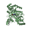
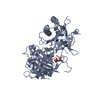
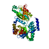
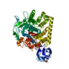
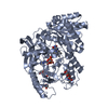
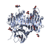
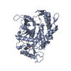
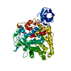
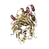
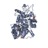
[English] 日本語
Yorodumi
- PDB-6f72: Crystal structure of VAO-type flavoprotein MtVAO615 at pH 7.5 fro... -

+
Open data


ID or keywords:

Loading...

-
Basic information

Entry
Database: PDB / ID: 6f72
TitleCrystal structure of VAO-type flavoprotein MtVAO615 at pH 7.5 from Myceliophthora thermophila C1
ComponentsMtVAO615
KeywordsFLAVOPROTEIN / VAO-type / FAD / oxidoreductase
Function / homology
Function and homology information


organic substance biosynthetic process / secondary metabolite biosynthetic process / FAD binding / oxidoreductase activity / extracellular region
Similarity search - Function
Berberine/berberine-like / Berberine and berberine like / FAD linked oxidase, N-terminal / FAD binding domain / FAD-binding domain, PCMH-type / PCMH-type FAD-binding domain profile. / FAD-binding, type PCMH, subdomain 2 / FAD-binding, type PCMH-like superfamily
Similarity search - Domain/homology
FLAVIN-ADENINE DINUCLEOTIDE / VAO-type flavoprotein oxidase VAO615
Similarity search - Component
Biological speciesMyceliophthora thermophila (fungus)
MethodX-RAY DIFFRACTION / MOLECULAR REPLACEMENT / Resolution: 2 Å
AuthorsRozeboom, H.J. / Fraaije, M.W.
CitationJournal: Molecules / Year: 2018
Title: Characterization of Two VAO-Type Flavoprotein Oxidases from Myceliophthora thermophila.
Authors: Ferrari, A.R. / Rozeboom, H.J. / Vugts, A.S.C. / Koetsier, M.J. / Floor, R. / Fraaije, M.W.
History
DepositionDec 7, 2017Deposition site: PDBE / Processing site: PDBE
Revision 1.0Jan 10, 2018Provider: repository / Type: Initial release
Revision 1.1Jan 17, 2018Group: Database references / Category: citation / citation_author
Item: _citation.country / _citation.journal_abbrev ..._citation.country / _citation.journal_abbrev / _citation.journal_id_CSD / _citation.journal_id_ISSN / _citation.journal_volume / _citation.pdbx_database_id_DOI / _citation.pdbx_database_id_PubMed / _citation.title / _citation.year / _citation_author.name
Revision 2.0Jul 29, 2020Group: Atomic model / Data collection ...Atomic model / Data collection / Derived calculations / Refinement description / Structure summary
Category: atom_site / chem_comp ...atom_site / chem_comp / entity / pdbx_branch_scheme / pdbx_chem_comp_identifier / pdbx_entity_branch / pdbx_entity_branch_descriptor / pdbx_entity_branch_link / pdbx_entity_branch_list / pdbx_entity_nonpoly / pdbx_nonpoly_scheme / pdbx_struct_assembly_gen / refine_hist / struct_asym / struct_conn / struct_site / struct_site_gen
Item: _atom_site.B_iso_or_equiv / _atom_site.Cartn_x ..._atom_site.B_iso_or_equiv / _atom_site.Cartn_x / _atom_site.Cartn_y / _atom_site.Cartn_z / _atom_site.auth_asym_id / _atom_site.auth_atom_id / _atom_site.auth_comp_id / _atom_site.auth_seq_id / _atom_site.label_asym_id / _atom_site.label_atom_id / _atom_site.label_comp_id / _atom_site.label_entity_id / _atom_site.type_symbol / _chem_comp.name / _chem_comp.type / _pdbx_struct_assembly_gen.asym_id_list / _refine_hist.d_res_low / _struct_conn.pdbx_role / _struct_conn.ptnr1_auth_asym_id / _struct_conn.ptnr1_auth_seq_id / _struct_conn.ptnr1_label_asym_id / _struct_conn.ptnr2_auth_asym_id / _struct_conn.ptnr2_auth_seq_id / _struct_conn.ptnr2_label_asym_id
Description: Carbohydrate remediation / Provider: repository / Type: Remediation

-
Structure visualization

Structure viewerMolecule:
MolmilJmol/JSmol

Downloads & links

-
Assembly

Deposited unit
A: MtVAO615
hetero molecules


Theoretical massNumber of molelcules
Total (without water)64,9098
Polymers62,0241
Non-polymers2,8857
Water6,593366
1


  • Idetical with deposited unit
  • defined by author&software
TypeNameSymmetry operationNumber
identity operation1_555x,y,z1
Buried area3700 Å2
ΔGint19 kcal/mol
Surface area21030 Å2
MethodPISA
Unit cell
Length a, b, c (Å)59.700, 100.930, 111.800
Angle α, β, γ (deg.)90.00, 90.00, 90.00
Int Tables number19
Space group name H-MP212121

-
Components

-
Protein , 1 types, 1 molecules A

#1: Protein MtVAO615


Mass: 62024.320 Da / Num. of mol.: 1
Source method: isolated from a genetically manipulated source
Details: First 17 residues are a signal sequence
Source: (gene. exp.) Myceliophthora thermophila (strain ATCC 42464 / BCRC 31852 / DSM 1799) (fungus)
Strain: ATCC 42464 / BCRC 31852 / DSM 1799 / Gene: MYCTH_2305637
Production host: Myceliophthora thermophila ATCC 42464 (fungus)
References: UniProt: G2QDQ9

-
Sugars , 3 types, 6 molecules

#2: Polysaccharide 2-acetamido-2-deoxy-beta-D-glucopyranose-(1-4)-2-acetamido-2-deoxy-beta-D-glucopyranose


Type: oligosaccharide / Mass: 424.401 Da / Num. of mol.: 2
Source method: isolated from a genetically manipulated source
DescriptorTypeProgram
DGlcpNAcb1-4DGlcpNAcb1-Glycam Condensed SequenceGMML 1.0
WURCS=2.0/1,2,1/[a2122h-1b_1-5_2*NCC/3=O]/1-1/a4-b1WURCSPDB2Glycan 1.1.0
[]{[(4+1)][b-D-GlcpNAc]{[(4+1)][b-D-GlcpNAc]{}}}LINUCSPDB-CARE
#3: Polysaccharide beta-D-mannopyranose-(1-4)-2-acetamido-2-deoxy-beta-D-glucopyranose-(1-4)-2-acetamido-2-deoxy-beta- ...beta-D-mannopyranose-(1-4)-2-acetamido-2-deoxy-beta-D-glucopyranose-(1-4)-2-acetamido-2-deoxy-beta-D-glucopyranose


Type: oligosaccharide / Mass: 586.542 Da / Num. of mol.: 1
Source method: isolated from a genetically manipulated source
DescriptorTypeProgram
DManpb1-4DGlcpNAcb1-4DGlcpNAcb1-Glycam Condensed SequenceGMML 1.0
WURCS=2.0/2,3,2/[a2122h-1b_1-5_2*NCC/3=O][a1122h-1b_1-5]/1-1-2/a4-b1_b4-c1WURCSPDB2Glycan 1.1.0
[]{[(4+1)][b-D-GlcpNAc]{[(4+1)][b-D-GlcpNAc]{[(4+1)][b-D-Manp]{}}}}LINUCSPDB-CARE
#5: Sugar ChemComp-NAG / 2-acetamido-2-deoxy-beta-D-glucopyranose / N-Acetylglucosamine


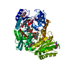
Type: D-saccharide, beta linking / Mass: 221.208 Da / Num. of mol.: 3
Source method: isolated from a genetically manipulated source
Formula: C8H15NO6
IdentifierTypeProgram
DGlcpNAcbCONDENSED IUPAC CARBOHYDRATE SYMBOLGMML 1.0
N-acetyl-b-D-glucopyranosamineCOMMON NAMEGMML 1.0
b-D-GlcpNAcIUPAC CARBOHYDRATE SYMBOLPDB-CARE 1.0
GlcNAcSNFG CARBOHYDRATE SYMBOLGMML 1.0

-
Non-polymers , 2 types, 367 molecules

#4: Chemical ChemComp-FAD / FLAVIN-ADENINE DINUCLEOTIDE / Flavin adenine dinucleotide


Mass: 785.550 Da / Num. of mol.: 1 / Source method: obtained synthetically / Formula: C27H33N9O15P2 / Comment: FAD*YM
#6: Water ChemComp-HOH / water / Water


Mass: 18.015 Da / Num. of mol.: 366 / Source method: isolated from a natural source / Formula: H2O

-
Experimental details

-
Experiment

ExperimentMethod: X-RAY DIFFRACTION / Number of used crystals: 1

-
Sample preparation

CrystalDensity Matthews: 2.7 Å3/Da / Density % sol: 54 %
Crystal growTemperature: 294 K / Method: vapor diffusion, hanging drop / Details: 11% PEG3350, 20% glycerol, 0.1 M HEPES pH 7.5

-
Data collection

DiffractionMean temperature: 110 K
Diffraction sourceSource: ROTATING ANODE / Type: BRUKER AXS MICROSTAR / Wavelength: 1.54 Å
DetectorType: MARRESEARCH / Detector: IMAGE PLATE / Date: Nov 29, 2014
RadiationMonochromator: Helios MX mirrors / Protocol: SINGLE WAVELENGTH / Monochromatic (M) / Laue (L): M / Scattering type: x-ray
Radiation wavelengthWavelength: 1.54 Å / Relative weight: 1
ReflectionResolution: 2→51.4 Å / Num. obs: 45720 / % possible obs: 98.1 % / Redundancy: 3.7 % / CC1/2: 0.988 / Rmerge(I) obs: 0.172 / Rpim(I) all: 0.114 / Net I/σ(I): 4.7
Reflection shellResolution: 2→2.05 Å / Redundancy: 3.2 % / Rmerge(I) obs: 0.986 / Mean I/σ(I) obs: 1 / Num. unique obs: 2754 / CC1/2: 0.333 / Rpim(I) all: 0.707 / % possible all: 81.1

-
Processing

Software
NameVersionClassification
REFMAC5.8.0155refinement
iMOSFLMdata reduction
Aimlessdata scaling
PHASERphasing
RefinementMethod to determine structure: MOLECULAR REPLACEMENT
Starting model: D_1200007842

Resolution: 2→51.4 Å / Cor.coef. Fo:Fc: 0.936 / Cor.coef. Fo:Fc free: 0.904 / Cross valid method: THROUGHOUT / ESU R: 0.212 / ESU R Free: 0.189 / Stereochemistry target values: MAXIMUM LIKELIHOOD / Details: HYDROGENS HAVE BEEN USED IF PRESENT IN THE INPUT
RfactorNum. reflection% reflectionSelection details
Rfree0.28044 2197 4.8 %RANDOM
Rwork0.2357 ---
obs0.23786 43454 98.06 %-
Solvent computationIon probe radii: 0.8 Å / Shrinkage radii: 0.8 Å / VDW probe radii: 1.2 Å / Solvent model: MASK
Displacement parametersBiso mean: 29.661 Å2
Baniso -1Baniso -2Baniso -3
1--0.68 Å2-0 Å2-0 Å2
2--0.18 Å2-0 Å2
3---0.5 Å2
Refinement stepCycle: 1 / Resolution: 2→51.4 Å
ProteinNucleic acidLigandSolventTotal
Num. atoms4188 0 190 366 4744
Refine LS restraints
Refine-IDTypeDev idealDev ideal targetNumber
X-RAY DIFFRACTIONr_bond_refined_d0.010.0194520
X-RAY DIFFRACTIONr_bond_other_d
X-RAY DIFFRACTIONr_angle_refined_deg1.4441.9856186
X-RAY DIFFRACTIONr_angle_other_deg
X-RAY DIFFRACTIONr_dihedral_angle_1_deg6.2985547
X-RAY DIFFRACTIONr_dihedral_angle_2_deg36.2423.526190
X-RAY DIFFRACTIONr_dihedral_angle_3_deg14.42115616
X-RAY DIFFRACTIONr_dihedral_angle_4_deg14.9221523
X-RAY DIFFRACTIONr_chiral_restr0.0920.2681
X-RAY DIFFRACTIONr_gen_planes_refined0.0060.0213476
X-RAY DIFFRACTIONr_gen_planes_other
X-RAY DIFFRACTIONr_nbd_refined
X-RAY DIFFRACTIONr_nbd_other
X-RAY DIFFRACTIONr_nbtor_refined
X-RAY DIFFRACTIONr_nbtor_other
X-RAY DIFFRACTIONr_xyhbond_nbd_refined
X-RAY DIFFRACTIONr_xyhbond_nbd_other
X-RAY DIFFRACTIONr_metal_ion_refined
X-RAY DIFFRACTIONr_metal_ion_other
X-RAY DIFFRACTIONr_symmetry_vdw_refined
X-RAY DIFFRACTIONr_symmetry_vdw_other
X-RAY DIFFRACTIONr_symmetry_hbond_refined
X-RAY DIFFRACTIONr_symmetry_hbond_other
X-RAY DIFFRACTIONr_symmetry_metal_ion_refined
X-RAY DIFFRACTIONr_symmetry_metal_ion_other
X-RAY DIFFRACTIONr_mcbond_it0.2270.9532191
X-RAY DIFFRACTIONr_mcbond_other
X-RAY DIFFRACTIONr_mcangle_it0.4121.4282737
X-RAY DIFFRACTIONr_mcangle_other
X-RAY DIFFRACTIONr_scbond_it0.8481.2232329
X-RAY DIFFRACTIONr_scbond_other
X-RAY DIFFRACTIONr_scangle_it
X-RAY DIFFRACTIONr_scangle_other
X-RAY DIFFRACTIONr_long_range_B_refined7.11915.9687390
X-RAY DIFFRACTIONr_long_range_B_other
X-RAY DIFFRACTIONr_rigid_bond_restr
X-RAY DIFFRACTIONr_sphericity_free
X-RAY DIFFRACTIONr_sphericity_bonded
LS refinement shellResolution: 1.998→2.05 Å / Total num. of bins used: 20
RfactorNum. reflection% reflection
Rfree0.376 136 -
Rwork0.399 2682 -
obs--82.64 %
Refinement TLS params.

Method: refined / Refine-ID: X-RAY DIFFRACTION

IDL112)L122)L132)L222)L232)L332)S11 (Å °)S12 (Å °)S13 (Å °)S21 (Å °)S22 (Å °)S23 (Å °)S31 (Å °)S32 (Å °)S33 (Å °)T112)T122)T132)T222)T232)T332)Origin x (Å)Origin y (Å)Origin z (Å)
10.3046-0.55960.07752.533-0.24111.709-0.03590.168-0.118-0.381-0.12920.26150.1835-0.05540.16520.6136-0.0685-0.00890.1188-0.07070.072221.986-19.9866.444
20.2167-0.0105-0.39541.2591-0.24970.793-0.06220.0191-0.0709-0.2956-0.0703-0.06750.2209-0.0050.13240.5669-0.00480.01880.0325-0.0190.039231.414-20.40914.815
30.94960.0072-0.0981.04390.0630.4640.01740.03360.0452-0.1492-0.0318-0.1135-0.0459-0.02780.01440.5011-0.00130.02840.02570.00480.016537.4452.64616.24
40.9894-0.04160.3990.6854-0.26571.5437-0.0017-0.04530.0330.1233-0.0267-0.23110.0830.23120.02850.54160.0243-0.03050.0537-0.00880.085756.569-3.69236.398
50.511-0.01190.02181.03540.08570.3764-0.0238-0.0721-0.02150.14110.0070.04360.01270.01150.01670.494600.02030.03030.00380.003631.728-7.47434.335
Refinement TLS group
IDRefine-IDRefine TLS-IDAuth asym-IDAuth seq-ID
1X-RAY DIFFRACTION1A27 - 66
2X-RAY DIFFRACTION2A67 - 162
3X-RAY DIFFRACTION3A163 - 294
4X-RAY DIFFRACTION4A295 - 406
5X-RAY DIFFRACTION5A407 - 574

+
About Yorodumi

-
News

-
Feb 9, 2022. New format data for meta-information of EMDB entries

New format data for meta-information of EMDB entries

  • Version 3 of the EMDB header file is now the official format.
  • The previous official version 1.9 will be removed from the archive.

Related info.:EMDB header

External links:wwPDB to switch to version 3 of the EMDB data model

-
Aug 12, 2020. Covid-19 info

Covid-19 info

URL: https://pdbj.org/emnavi/covid19.php

New page: Covid-19 featured information page in EM Navigator.

Related info.:Covid-19 info / Mar 5, 2020. Novel coronavirus structure data

+
Mar 5, 2020. Novel coronavirus structure data

Novel coronavirus structure data

Related info.:Yorodumi Speices / Aug 12, 2020. Covid-19 info

External links:COVID-19 featured content - PDBj / Molecule of the Month (242):Coronavirus Proteases

+
Jan 31, 2019. EMDB accession codes are about to change! (news from PDBe EMDB page)

EMDB accession codes are about to change! (news from PDBe EMDB page)

  • The allocation of 4 digits for EMDB accession codes will soon come to an end. Whilst these codes will remain in use, new EMDB accession codes will include an additional digit and will expand incrementally as the available range of codes is exhausted. The current 4-digit format prefixed with “EMD-” (i.e. EMD-XXXX) will advance to a 5-digit format (i.e. EMD-XXXXX), and so on. It is currently estimated that the 4-digit codes will be depleted around Spring 2019, at which point the 5-digit format will come into force.
  • The EM Navigator/Yorodumi systems omit the EMD- prefix.

Related info.:Q: What is EMD? / ID/Accession-code notation in Yorodumi/EM Navigator

External links:EMDB Accession Codes are Changing Soon! / Contact to PDBj

+
Jul 12, 2017. Major update of PDB

Major update of PDB

  • wwPDB released updated PDB data conforming to the new PDBx/mmCIF dictionary.
  • This is a major update changing the version number from 4 to 5, and with Remediation, in which all the entries are updated.
  • In this update, many items about electron microscopy experimental information are reorganized (e.g. em_software).
  • Now, EM Navigator and Yorodumi are based on the updated data.

External links:wwPDB Remediation / Enriched Model Files Conforming to OneDep Data Standards Now Available in the PDB FTP Archive

-
Yorodumi

Thousand views of thousand structures

  • Yorodumi is a browser for structure data from EMDB, PDB, SASBDB, etc.
  • This page is also the successor to EM Navigator detail page, and also detail information page/front-end page for Omokage search.
  • The word "yorodu" (or yorozu) is an old Japanese word meaning "ten thousand". "mi" (miru) is to see.

Related info.:EMDB / PDB / SASBDB / Comparison of 3 databanks / Yorodumi Search / Aug 31, 2016. New EM Navigator & Yorodumi / Yorodumi Papers / Jmol/JSmol / Function and homology information / Changes in new EM Navigator and Yorodumi

Read more