 Movie
Movie Controller

[English] 日本語
 Yorodumi
- PDB-6ezm: Imidazoleglycerol-phosphate dehydratase from Saccharomyces cerevisiae -
+ Open data

- Basic information
| Entry | Database: PDB / ID: 6ezm | ||||||||||||
|---|---|---|---|---|---|---|---|---|---|---|---|---|---|
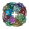
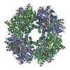
| Title | Imidazoleglycerol-phosphate dehydratase from Saccharomyces cerevisiae | ||||||||||||
 Components Components | Imidazoleglycerol-phosphate dehydratase | ||||||||||||
 Keywords Keywords | LYASE / Inhibitor / histidine biosynthesis / complex | ||||||||||||
| Function / homology |  Function and homology information Function and homology informationimidazoleglycerol-phosphate dehydratase / imidazoleglycerol-phosphate dehydratase activity / L-histidine biosynthetic process / metal ion binding Similarity search - Function | ||||||||||||
| Biological species |  | ||||||||||||
| Method | ELECTRON MICROSCOPY / single particle reconstruction / cryo EM / Resolution: 3.2 Å | ||||||||||||
 Authors Authors | Rawson, S. / Bisson, C. / Hurdiss, D.L. / Muench, S.P. | ||||||||||||
| Funding support |  United Kingdom, 3items United Kingdom, 3items
| ||||||||||||
 Citation Citation | Journal: Proc Natl Acad Sci U S A / Year: 2018 Title: Elucidating the structural basis for differing enzyme inhibitor potency by cryo-EM. Authors: Shaun Rawson / Claudine Bisson / Daniel L Hurdiss / Asif Fazal / Martin J McPhillie / Svetlana E Sedelnikova / Patrick J Baker / David W Rice / Stephen P Muench / Abstract: Histidine biosynthesis is an essential process in plants and microorganisms, making it an attractive target for the development of herbicides and antibacterial agents. Imidazoleglycerol-phosphate ...Histidine biosynthesis is an essential process in plants and microorganisms, making it an attractive target for the development of herbicides and antibacterial agents. Imidazoleglycerol-phosphate dehydratase (IGPD), a key enzyme within this pathway, has been biochemically characterized in both (IGPD) and (IGPD). The plant enzyme, having been the focus of in-depth structural analysis as part of an inhibitor development program, has revealed details about the reaction mechanism of IGPD, whereas the yeast enzyme has proven intractable to crystallography studies. The structure-activity relationship of potent triazole-phosphonate inhibitors of IGPD has been determined in both homologs, revealing that the lead inhibitor (C348) is an order of magnitude more potent against IGPD than IGPD; however, the molecular basis of this difference has not been established. Here we have used single-particle electron microscopy (EM) to study structural differences between the and IGPD homologs, which could influence the difference in inhibitor potency. The resulting EM maps at ∼3 Å are sufficient to de novo build the protein structure and identify the inhibitor binding site, which has been validated against the crystal structure of the IGPD/C348 complex. The structure of _IGPD reveals that a 24-amino acid insertion forms an extended loop region on the enzyme surface that lies adjacent to the active site, forming interactions with the substrate/inhibitor binding loop that may influence inhibitor potency. Overall, this study provides insights into the IGPD family and demonstrates the power of using an EM approach to study inhibitor binding. | ||||||||||||
| History |
|
- Structure visualization
| Movie |
Movie viewer |
|---|---|
| Structure viewer | Molecule: MolmilJmol/JSmol |
- Downloads & links
-Download
| PDBx/mmCIF format | 6ezm.cif.gz | 830.5 KB | Display | PDBx/mmCIF format |
|---|---|---|---|---|
| PDB format | pdb6ezm.ent.gz | 694.5 KB | Display | PDB format |
| PDBx/mmJSON format | 6ezm.json.gz | Tree view | PDBx/mmJSON format | |
| Others |  Other downloads Other downloads |
-Validation report
| Arichive directory | https://data.pdbj.org/pub/pdb/validation_reports/ez/6ezmftp://data.pdbj.org/pub/pdb/validation_reports/ez/6ezm | HTTPS FTP |
|---|
-Related structure data
| Related structure data | 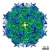 4160MC  3999C  6ezjC C: citing same article ( M: map data used to model this data |
|---|---|
| Similar structure data |


-Links
 PDBj
PDBj- Assembly
| Deposited unit | 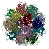
|
|---|---|
| 1 |
|
-Components
| #1: Protein | Mass: 23867.197 Da / Num. of mol.: 24 Source method: isolated from a genetically manipulated source Source: (gene. exp.) Strain: ATCC 204508 / S288c / Gene: HIS3, YOR202W / Production host:  References: UniProt: P06633, imidazoleglycerol-phosphate dehydratase #2: Chemical | ChemComp-MN /   Mass: 54.938 Da / Num. of mol.: 48 / Source method: obtained synthetically / Formula: Mn Mass: 54.938 Da / Num. of mol.: 48 / Source method: obtained synthetically / Formula: Mn#3: Chemical | ChemComp-5LD / [(   Mass: 207.124 Da / Num. of mol.: 24 / Source method: obtained synthetically / Formula: C5H10N3O4P / Feature type: SUBJECT OF INVESTIGATION Mass: 207.124 Da / Num. of mol.: 24 / Source method: obtained synthetically / Formula: C5H10N3O4P / Feature type: SUBJECT OF INVESTIGATION |
|---|
-Experimental details
-Experiment
| Experiment | Method: ELECTRON MICROSCOPY |
|---|---|
| EM experiment | Aggregation state: PARTICLE / 3D reconstruction method: single particle reconstruction |
- Sample preparation
| Component | Name: Imidazoleglycerol-phosphate dehydratase (IGPD) in complex with triazole-phosphonate inhibitor (C384) Type: COMPLEX / Entity ID: #1 / Source: RECOMBINANT |
|---|---|
| Molecular weight | Value: 0.57 MDa / Experimental value: YES |
| Source (natural) | Organism: |
| Source (recombinant) | Organism: |
| Buffer solution | pH: 7.5 |
| Specimen | Conc.: 0.5 mg/ml / Embedding applied: NO / Shadowing applied: NO / Staining applied: NO / Vitrification applied: YES |
| Specimen support | Grid material: COPPER / Grid type: Quantifoil R2/2 |
| Vitrification | Instrument: FEI VITROBOT MARK IV / Cryogen name: ETHANE / Humidity: 100 % |
- Electron microscopy imaging
| Experimental equipment |  Model: Titan Krios / Image courtesy: FEI Company |
|---|---|
| Microscopy | Model: FEI TITAN KRIOS |
| Electron gun | Electron source:  FIELD EMISSION GUN / Accelerating voltage: 300 kV / Illumination mode: FLOOD BEAM FIELD EMISSION GUN / Accelerating voltage: 300 kV / Illumination mode: FLOOD BEAM |
| Electron lens | Mode: BRIGHT FIELD |
| Specimen holder | Cryogen: NITROGEN / Specimen holder model: FEI TITAN KRIOS AUTOGRID HOLDER |
| Image recording | Electron dose: 1.3 e/Å2 / Detector mode: INTEGRATING / Film or detector model: FEI FALCON III (4k x 4k) / Num. of grids imaged: 1 / Num. of real images: 2827 |
| Image scans | Movie frames/image: 39 |
- Processing
| Software | Name: PHENIX / Version: 1.11.1_2575: / Classification: refinement | ||||||||||||||||||||||||
|---|---|---|---|---|---|---|---|---|---|---|---|---|---|---|---|---|---|---|---|---|---|---|---|---|---|
| EM software |
| ||||||||||||||||||||||||
| CTF correction | Type: PHASE FLIPPING AND AMPLITUDE CORRECTION | ||||||||||||||||||||||||
| Particle selection | Num. of particles selected: 365498 | ||||||||||||||||||||||||
| Symmetry | Point symmetry: O (octahedral) | ||||||||||||||||||||||||
| 3D reconstruction | Resolution: 3.2 Å / Resolution method: FSC 0.143 CUT-OFF / Num. of particles: 11560 / Details: Relion2.1 auto-refine / Num. of class averages: 1 / Symmetry type: POINT | ||||||||||||||||||||||||
| Refine LS restraints |
|
