 Movie
Movie Controller


+ Open data

- Basic information
| Entry | Database: PDB / ID: 6ez7 | ||||||
|---|---|---|---|---|---|---|---|
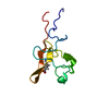
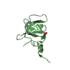
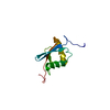
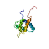
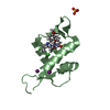
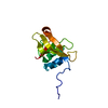
| Title | Pes4 RRM3 Structure | ||||||
 Components Components | Protein PES4 | ||||||
 Keywords Keywords | RNA BINDING PROTEIN / RNA Recognition Motif (RRM) | ||||||
| Function / homology |  Function and homology information Function and homology informationmRNA localization resulting in post-transcriptional regulation of gene expression / Deadenylation of mRNA / sporulation / prospore membrane / mRNA metabolic process / poly(A) binding / poly(U) RNA binding / Translation initiation complex formation / Nonsense Mediated Decay (NMD) independent of the Exon Junction Complex (EJC) / Nonsense Mediated Decay (NMD) enhanced by the Exon Junction Complex (EJC) ...mRNA localization resulting in post-transcriptional regulation of gene expression / Deadenylation of mRNA / sporulation / prospore membrane / mRNA metabolic process / poly(A) binding / poly(U) RNA binding / Translation initiation complex formation / Nonsense Mediated Decay (NMD) independent of the Exon Junction Complex (EJC) / Nonsense Mediated Decay (NMD) enhanced by the Exon Junction Complex (EJC) / L13a-mediated translational silencing of Ceruloplasmin expression / mRNA 3'-UTR binding / cytoplasmic stress granule / regulation of translation / ribonucleoprotein complex / nucleus / cytosol Similarity search - Function | ||||||
| Biological species |  | ||||||
| Method |  X-RAY DIFFRACTION / SYNCHROTRON / MOLECULAR REPLACEMENT / Resolution: 1.9 Å X-RAY DIFFRACTION / SYNCHROTRON / MOLECULAR REPLACEMENT / Resolution: 1.9 Å | ||||||
 Authors Authors | Mohamad, N. / Bravo, J. | ||||||
| Funding support |  Spain, 1items Spain, 1items
| ||||||
 Citation Citation | Journal: To Be Published Title: Pes4 RRM3 Structure Authors: Mohamad, N. / Bravo, J. | ||||||
| History |
|
- Structure visualization
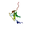
| Structure viewer | Molecule: MolmilJmol/JSmol |
|---|
- Downloads & links
-Download
| PDBx/mmCIF format | 6ez7.cif.gz | 48.8 KB | Display | PDBx/mmCIF format |
|---|---|---|---|---|
| PDB format | pdb6ez7.ent.gz | 34.4 KB | Display | PDB format |
| PDBx/mmJSON format | 6ez7.json.gz | Tree view | PDBx/mmJSON format | |
| Others |  Other downloads Other downloads |
-Validation report
| Arichive directory | https://data.pdbj.org/pub/pdb/validation_reports/ez/6ez7ftp://data.pdbj.org/pub/pdb/validation_reports/ez/6ez7 | HTTPS FTP |
|---|
-Related structure data
| Related structure data |  5d77S S: Starting model for refinement |
|---|---|
| Similar structure data |

-Links
 PDBj
PDBj


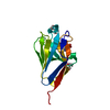
- Assembly
| Deposited unit | 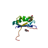
| ||||||||
|---|---|---|---|---|---|---|---|---|---|
| 1 |
| ||||||||
| Unit cell |
| ||||||||
| Components on special symmetry positions |
|
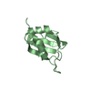
-Components
| #1: Protein | Mass: 9763.218 Da / Num. of mol.: 1 Source method: isolated from a genetically manipulated source Source: (gene. exp.)  | ||||
|---|---|---|---|---|---|
| #2: Chemical |   Mass: 106.120 Da / Num. of mol.: 2 / Source method: obtained synthetically / Formula: C4H10O3 Mass: 106.120 Da / Num. of mol.: 2 / Source method: obtained synthetically / Formula: C4H10O3#3: Water | ChemComp-HOH / |  Mass: 18.015 Da / Num. of mol.: 46 / Source method: isolated from a natural source / Formula: H2O Mass: 18.015 Da / Num. of mol.: 46 / Source method: isolated from a natural source / Formula: H2OSequence details | protein used to set for the crystallization plates was Pes4(303-473) however, the part that ...protein used to set for the crystallization plates was Pes4(303-473) however, the part that crystallized was Pes4(303-384) | |
-Experimental details
-Experiment
| Experiment | Method: X-RAY DIFFRACTION / Number of used crystals: 1 |
|---|
- Sample preparation
| Crystal | Density Matthews: 3.24 Å3/Da / Density % sol: 62.08 % |
|---|---|
| Crystal grow | Temperature: 294 K / Method: vapor diffusion, sitting drop / pH: 7.4 Details: 200 mM Ammonium Sulfate, 30% PEG 8000, 1M MES/NaOH pH 7.4 (additive) |
-Data collection
| Diffraction | Mean temperature: 100 K |
|---|---|
| Diffraction source | Source: SYNCHROTRON / Site: Diamond  / Beamline: I03 / Wavelength: 0.97623 Å / Beamline: I03 / Wavelength: 0.97623 Å |
| Detector | Type: DECTRIS PILATUS 6M / Detector: PIXEL / Date: Sep 17, 2016 |
| Radiation | Protocol: SINGLE WAVELENGTH / Monochromatic (M) / Laue (L): M / Scattering type: x-ray |
| Radiation wavelength | Wavelength: 0.97623 Å / Relative weight: 1 |
| Reflection | Resolution: 1.9→29.212 Å / Num. obs: 10165 / % possible obs: 99.99 % / Redundancy: 13.3 % / Rmerge(I) obs: 0.2455 / Rrim(I) all: 0.2546 / Net I/σ(I): 6.63 |
| Reflection shell | Resolution: 1.9→1.94 Å / % possible all: 100 |
- Processing
| Software |
| ||||||||||||||||||||||||||||||||||||||||||||||||||||||||||||||||||||||||||||||||||||||||||||||||||||
|---|---|---|---|---|---|---|---|---|---|---|---|---|---|---|---|---|---|---|---|---|---|---|---|---|---|---|---|---|---|---|---|---|---|---|---|---|---|---|---|---|---|---|---|---|---|---|---|---|---|---|---|---|---|---|---|---|---|---|---|---|---|---|---|---|---|---|---|---|---|---|---|---|---|---|---|---|---|---|---|---|---|---|---|---|---|---|---|---|---|---|---|---|---|---|---|---|---|---|---|---|---|
| Refinement | Method to determine structure: MOLECULAR REPLACEMENT Starting model: 5D77 Resolution: 1.9→29.212 Å / SU ML: 0.2 / Cross valid method: FREE R-VALUE / σ(F): 0 / Phase error: 29.48
| ||||||||||||||||||||||||||||||||||||||||||||||||||||||||||||||||||||||||||||||||||||||||||||||||||||
| Solvent computation | Shrinkage radii: 0.9 Å / VDW probe radii: 1.11 Å | ||||||||||||||||||||||||||||||||||||||||||||||||||||||||||||||||||||||||||||||||||||||||||||||||||||
| Refinement step | Cycle: LAST / Resolution: 1.9→29.212 Å
| ||||||||||||||||||||||||||||||||||||||||||||||||||||||||||||||||||||||||||||||||||||||||||||||||||||
| Refine LS restraints |
| ||||||||||||||||||||||||||||||||||||||||||||||||||||||||||||||||||||||||||||||||||||||||||||||||||||
| LS refinement shell |
| ||||||||||||||||||||||||||||||||||||||||||||||||||||||||||||||||||||||||||||||||||||||||||||||||||||
| Refinement TLS params. | Method: refined / Refine-ID: X-RAY DIFFRACTION
| ||||||||||||||||||||||||||||||||||||||||||||||||||||||||||||||||||||||||||||||||||||||||||||||||||||
| Refinement TLS group |
|
