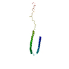
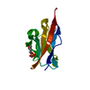
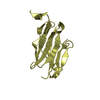
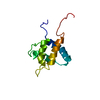
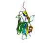
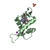
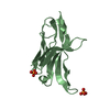
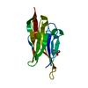
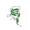
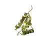
[English] 日本語
Yorodumi
- PDB-2x6m: Structure of a single domain camelid antibody fragment in complex... -

+
Open data


ID or keywords:

Loading...

-
Basic information

Entry
Database: PDB / ID: 2x6m
TitleStructure of a single domain camelid antibody fragment in complex with a C-terminal peptide of alpha-synuclein
Components
  • ALPHA-SYNUCLEIN PEPTIDE
  • HEAVY CHAIN VARIABLE DOMAIN FROM DROMEDARY
KeywordsIMMUNE SYSTEM / PARKINSON'S DISEASE / ALZHEIMER DISEASE AMYLOID / NANOBODY / AFFINITY TAG
Function / homology
Function and homology information


negative regulation of mitochondrial electron transport, NADH to ubiquinone / neutral lipid metabolic process / regulation of phospholipase activity / negative regulation of monooxygenase activity / regulation of acyl-CoA biosynthetic process / negative regulation of dopamine uptake involved in synaptic transmission / negative regulation of norepinephrine uptake / positive regulation of glutathione peroxidase activity / positive regulation of SNARE complex assembly / positive regulation of hydrogen peroxide catabolic process ...negative regulation of mitochondrial electron transport, NADH to ubiquinone / neutral lipid metabolic process / regulation of phospholipase activity / negative regulation of monooxygenase activity / regulation of acyl-CoA biosynthetic process / negative regulation of dopamine uptake involved in synaptic transmission / negative regulation of norepinephrine uptake / positive regulation of glutathione peroxidase activity / positive regulation of SNARE complex assembly / positive regulation of hydrogen peroxide catabolic process / supramolecular fiber / negative regulation of transporter activity / mitochondrial membrane organization / negative regulation of chaperone-mediated autophagy / regulation of reactive oxygen species biosynthetic process / positive regulation of protein localization to cell periphery / regulation of synaptic vesicle recycling / negative regulation of platelet-derived growth factor receptor signaling pathway / negative regulation of exocytosis / regulation of glutamate secretion / regulation of norepinephrine uptake / response to iron(II) ion / SNARE complex assembly / positive regulation of neurotransmitter secretion / dopamine biosynthetic process / regulation of locomotion / positive regulation of inositol phosphate biosynthetic process / synaptic vesicle priming / regulation of macrophage activation / negative regulation of microtubule polymerization / dopamine uptake involved in synaptic transmission / synaptic vesicle transport / dynein complex binding / positive regulation of receptor recycling / regulation of dopamine secretion / protein kinase inhibitor activity / negative regulation of thrombin-activated receptor signaling pathway / response to type II interferon / cuprous ion binding / positive regulation of exocytosis / synaptic vesicle exocytosis / kinesin binding / positive regulation of endocytosis / mitochondrial ATP synthesis coupled electron transport / cysteine-type endopeptidase inhibitor activity involved in apoptotic process / response to magnesium ion / regulation of presynapse assembly / synaptic vesicle endocytosis / negative regulation of serotonin uptake / alpha-tubulin binding / localization / phospholipid metabolic process / supramolecular fiber organization / axon terminus / inclusion body / cellular response to copper ion / cellular response to epinephrine stimulus / Hsp70 protein binding / excitatory postsynaptic potential / response to interleukin-1 / adult locomotory behavior / SNARE binding / positive regulation of release of sequestered calcium ion into cytosol / fatty acid metabolic process / long-term synaptic potentiation / phosphoprotein binding / protein tetramerization / regulation of transmembrane transporter activity / synapse organization / regulation of long-term neuronal synaptic plasticity / microglial cell activation / negative regulation of protein kinase activity / protein destabilization / negative regulation of cysteine-type endopeptidase activity involved in apoptotic process / ferrous iron binding / tau protein binding / PKR-mediated signaling / positive regulation of protein serine/threonine kinase activity / receptor internalization / phospholipid binding / synaptic vesicle membrane / positive regulation of inflammatory response / activation of cysteine-type endopeptidase activity involved in apoptotic process / actin cytoskeleton / positive regulation of peptidyl-serine phosphorylation / actin binding / cell cortex / histone binding / cellular response to oxidative stress / growth cone / postsynapse / chemical synaptic transmission / neuron apoptotic process / negative regulation of neuron apoptotic process / amyloid fibril formation / response to lipopolysaccharide / molecular adaptor activity / oxidoreductase activity / lysosome / transcription cis-regulatory region binding
Similarity search - Function
Synuclein / Alpha-synuclein / Synuclein / Immunoglobulins / Immunoglobulin-like / Sandwich / Mainly Beta
Similarity search - Domain/homology
Biological speciesCAMELUS DROMEDARIUS (Arabian camel)
HOMO SAPIENS (human)
MethodX-RAY DIFFRACTION / SYNCHROTRON / MOLECULAR REPLACEMENT / Resolution: 1.62 Å
AuthorsDeGenst, E. / Guilliams, T. / Wellens, J. / O'Day, E.M. / Waudby, C.A. / Meehan, S. / Dumoulin, M. / Hsu, S.-T.D. / Cremades, N. / Verschueren, K.H.G. ...DeGenst, E. / Guilliams, T. / Wellens, J. / O'Day, E.M. / Waudby, C.A. / Meehan, S. / Dumoulin, M. / Hsu, S.-T.D. / Cremades, N. / Verschueren, K.H.G. / Pardon, E. / Wyns, L. / Steyaert, J. / Christodoulou, J. / Dobson, C.M.
CitationJournal: J.Mol.Biol. / Year: 2010
Title: Structure and Properties of a Complex of Alpha-Synuclein and a Single-Domain Camelid Antibody.
Authors: De Genst, E.J. / Guilliams, T. / Wellens, J. / O'Day, E.M. / Waudby, C.A. / Meehan, S. / Dumoulin, M. / Hsu, S.-T.D. / Cremades, N. / Verschueren, K.H.G. / Pardon, E. / Wyns, L. / Steyaert, ...Authors: De Genst, E.J. / Guilliams, T. / Wellens, J. / O'Day, E.M. / Waudby, C.A. / Meehan, S. / Dumoulin, M. / Hsu, S.-T.D. / Cremades, N. / Verschueren, K.H.G. / Pardon, E. / Wyns, L. / Steyaert, J. / Christodoulou, J. / Dobson, C.M.
History
DepositionFeb 18, 2010Deposition site: PDBE / Processing site: PDBE
Revision 1.0Jun 23, 2010Provider: repository / Type: Initial release
Revision 1.1Aug 24, 2011Group: Database references / Structure summary / Version format compliance
Revision 1.2Mar 29, 2017Group: Other
Revision 1.3Jul 24, 2019Group: Data collection / Category: diffrn_source / Item: _diffrn_source.pdbx_synchrotron_site
Revision 1.4Dec 20, 2023Group: Data collection / Database references ...Data collection / Database references / Other / Refinement description
Category: chem_comp_atom / chem_comp_bond ...chem_comp_atom / chem_comp_bond / database_2 / pdbx_database_status / pdbx_initial_refinement_model
Item: _database_2.pdbx_DOI / _database_2.pdbx_database_accession / _pdbx_database_status.status_code_sf

-
Structure visualization

Structure viewerMolecule:
MolmilJmol/JSmol

Downloads & links

-
Assembly

Deposited unit
A: HEAVY CHAIN VARIABLE DOMAIN FROM DROMEDARY
B: ALPHA-SYNUCLEIN PEPTIDE


Theoretical massNumber of molelcules
Total (without water)14,5092
Polymers14,5092
Non-polymers00
Water3,747208
1


  • Idetical with deposited unit
  • defined by author&software
TypeNameSymmetry operationNumber
identity operation1_555x,y,z1
Buried area620 Å2
ΔGint-0.8 kcal/mol
Surface area6540 Å2
MethodPISA
Unit cell
Length a, b, c (Å)60.170, 63.120, 62.870
Angle α, β, γ (deg.)90.00, 90.00, 90.00
Int Tables number20
Space group name H-MC2221
Components on special symmetry positions
IDModelComponents
11A-2020-

HOH

21A-2078-

HOH

-
Components

#1: Antibody HEAVY CHAIN VARIABLE DOMAIN FROM DROMEDARY


Mass: 13437.815 Da / Num. of mol.: 1
Source method: isolated from a genetically manipulated source
Details: ENGINEERED FROM IMMUNISED DROMEDARY / Source: (gene. exp.) CAMELUS DROMEDARIUS (Arabian camel) / Tissue: BLOOD / Cell: LYMPHOCYTE / Production host: ESCHERICHIA COLI (E. coli) / Strain (production host): WK6
#2: Protein/peptide ALPHA-SYNUCLEIN PEPTIDE


Mass: 1071.052 Da / Num. of mol.: 1
Fragment: C-TERMINAL FRAGMENT OF ALPHA-SYNUCLEIN, RESIDUES 132-140
Source method: obtained synthetically / Source: (synth.) HOMO SAPIENS (human) / References: UniProt: P37840
#3: Water ChemComp-HOH / water


Mass: 18.015 Da / Num. of mol.: 208 / Source method: isolated from a natural source / Formula: H2O

-
Experimental details

-
Experiment

ExperimentMethod: X-RAY DIFFRACTION / Number of used crystals: 1

-
Sample preparation

CrystalDensity Matthews: 2.01 Å3/Da / Density % sol: 38.84 % / Description: NONE
Crystal growpH: 7.5 / Details: 25% PEG6000, 100 MM HEPES PH 7.5, 100 MM LICL

-
Data collection

DiffractionMean temperature: 173 K
Diffraction sourceSource: SYNCHROTRON / Site: EMBL/DESY, HAMBURG / Beamline: X11 / Wavelength: 0.815
DetectorType: MAR555 FLAT PANEL / Detector: IMAGE PLATE
RadiationMonochromator: SI(111) / Protocol: SINGLE WAVELENGTH / Monochromatic (M) / Laue (L): M / Scattering type: x-ray
Radiation wavelengthWavelength: 0.815 Å / Relative weight: 1
ReflectionResolution: 1.62→30 Å / Num. obs: 15482 / % possible obs: 99.9 % / Observed criterion σ(I): 0 / Redundancy: 14.3 % / Biso Wilson estimate: 18.126 Å2 / Rmerge(I) obs: 0.12 / Net I/σ(I): 23.3
Reflection shellResolution: 1.62→1.71 Å / Redundancy: 14.2 % / Rmerge(I) obs: 0.54 / Mean I/σ(I) obs: 4.3 / % possible all: 99.9

-
Processing

Software
NameVersionClassification
REFMAC5.5.0102refinement
MOSFLMdata reduction
SCALAdata scaling
PHASERphasing
RefinementMethod to determine structure: MOLECULAR REPLACEMENT
Starting model: PDB ENTRY 1HCV
Resolution: 1.62→43.56 Å / Cor.coef. Fo:Fc: 0.97 / Cor.coef. Fo:Fc free: 0.95 / SU B: 3.577 / SU ML: 0.056 / Cross valid method: THROUGHOUT / ESU R: 0.094 / ESU R Free: 0.099 / Stereochemistry target values: MAXIMUM LIKELIHOOD
Details: HYDROGENS HAVE BEEN ADDED IN THE RIDING POSITIONS.U VALUES RESIDUAL ONLY. ATOM RECORD CONTAINS SUM OF TLS AND RESIDUAL B FACTORS.ANISOU RECORD CONTAINS SUM OF TLS AND RESIDUAL U FACTORS.
RfactorNum. reflection% reflectionSelection details
Rfree0.20258 1101 7.1 %RANDOM
Rwork0.15634 ---
obs0.1596 14361 99.93 %-
Solvent computationIon probe radii: 0.8 Å / Shrinkage radii: 0.8 Å / VDW probe radii: 1.4 Å / Solvent model: BABINET MODEL WITH MASK
Displacement parametersBiso mean: 17.91 Å2
Baniso -1Baniso -2Baniso -3
1--0.47 Å20 Å20 Å2
2---0.19 Å20 Å2
3---0.66 Å2
Refinement stepCycle: LAST / Resolution: 1.62→43.56 Å
ProteinNucleic acidLigandSolventTotal
Num. atoms993 0 0 208 1201
Refine LS restraints
Refine-IDTypeDev idealDev ideal targetNumber
X-RAY DIFFRACTIONr_bond_refined_d0.0180.0211060
X-RAY DIFFRACTIONr_bond_other_d
X-RAY DIFFRACTIONr_angle_refined_deg1.6931.9381446
X-RAY DIFFRACTIONr_angle_other_deg
X-RAY DIFFRACTIONr_dihedral_angle_1_deg7.0315147
X-RAY DIFFRACTIONr_dihedral_angle_2_deg33.15423.87849
X-RAY DIFFRACTIONr_dihedral_angle_3_deg12.93115166
X-RAY DIFFRACTIONr_dihedral_angle_4_deg24.031157
X-RAY DIFFRACTIONr_chiral_restr0.1470.2146
X-RAY DIFFRACTIONr_gen_planes_refined0.0150.021843
X-RAY DIFFRACTIONr_gen_planes_other
X-RAY DIFFRACTIONr_nbd_refined
X-RAY DIFFRACTIONr_nbd_other
X-RAY DIFFRACTIONr_nbtor_refined
X-RAY DIFFRACTIONr_nbtor_other
X-RAY DIFFRACTIONr_xyhbond_nbd_refined
X-RAY DIFFRACTIONr_xyhbond_nbd_other
X-RAY DIFFRACTIONr_metal_ion_refined
X-RAY DIFFRACTIONr_metal_ion_other
X-RAY DIFFRACTIONr_symmetry_vdw_refined
X-RAY DIFFRACTIONr_symmetry_vdw_other
X-RAY DIFFRACTIONr_symmetry_hbond_refined
X-RAY DIFFRACTIONr_symmetry_hbond_other
X-RAY DIFFRACTIONr_symmetry_metal_ion_refined
X-RAY DIFFRACTIONr_symmetry_metal_ion_other
X-RAY DIFFRACTIONr_mcbond_it1.5311.5671
X-RAY DIFFRACTIONr_mcbond_other
X-RAY DIFFRACTIONr_mcangle_it2.48121068
X-RAY DIFFRACTIONr_mcangle_other
X-RAY DIFFRACTIONr_scbond_it3.5613389
X-RAY DIFFRACTIONr_scbond_other
X-RAY DIFFRACTIONr_scangle_it5.5394.5369
X-RAY DIFFRACTIONr_scangle_other
X-RAY DIFFRACTIONr_long_range_B_refined
X-RAY DIFFRACTIONr_long_range_B_other
X-RAY DIFFRACTIONr_rigid_bond_restr
X-RAY DIFFRACTIONr_sphericity_free
X-RAY DIFFRACTIONr_sphericity_bonded
LS refinement shellResolution: 1.623→1.665 Å / Total num. of bins used: 20
RfactorNum. reflection% reflection
Rfree0.458 87 -
Rwork0.318 1032 -
obs--99.73 %
Refinement TLS params.

Method: refined / Refine-ID: X-RAY DIFFRACTION

IDL112)L122)L132)L222)L232)L332)S11 (Å °)S12 (Å °)S13 (Å °)S21 (Å °)S22 (Å °)S23 (Å °)S31 (Å °)S32 (Å °)S33 (Å °)T112)T122)T132)T222)T232)T332)Origin x (Å)Origin y (Å)Origin z (Å)
10.7485-0.26390.45410.6799-0.29910.6967-0.01060.02520.02360.0086-0.02890.02360.0076-0.01190.03950.0291-0.0045-0.00790.0268-0.00620.0243-15.045914.95079.2345
217.2343-0.81233.8938-1.2996-0.54270.7132-0.0793-0.1768-0.25080.05980.03640.04540.0898-0.14310.04290.0004-0.0326-0.01210.0872-0.00850.0791-34.658815.21963.5248
Refinement TLS group
IDRefine-IDRefine TLS-IDAuth asym-IDAuth seq-ID
1X-RAY DIFFRACTION1A2 - 127
2X-RAY DIFFRACTION2B4 - 9

+
About Yorodumi

-
News

-
Feb 9, 2022. New format data for meta-information of EMDB entries

New format data for meta-information of EMDB entries

  • Version 3 of the EMDB header file is now the official format.
  • The previous official version 1.9 will be removed from the archive.

Related info.:EMDB header

External links:wwPDB to switch to version 3 of the EMDB data model

-
Aug 12, 2020. Covid-19 info

Covid-19 info

URL: https://pdbjlvh1.pdbj.org/emnavi/covid19.php

New page: Covid-19 featured information page in EM Navigator.

Related info.:Covid-19 info / Mar 5, 2020. Novel coronavirus structure data

+
Mar 5, 2020. Novel coronavirus structure data

Novel coronavirus structure data

Related info.:Yorodumi Speices / Aug 12, 2020. Covid-19 info

External links:COVID-19 featured content - PDBj / Molecule of the Month (242):Coronavirus Proteases

+
Jan 31, 2019. EMDB accession codes are about to change! (news from PDBe EMDB page)

EMDB accession codes are about to change! (news from PDBe EMDB page)

  • The allocation of 4 digits for EMDB accession codes will soon come to an end. Whilst these codes will remain in use, new EMDB accession codes will include an additional digit and will expand incrementally as the available range of codes is exhausted. The current 4-digit format prefixed with “EMD-” (i.e. EMD-XXXX) will advance to a 5-digit format (i.e. EMD-XXXXX), and so on. It is currently estimated that the 4-digit codes will be depleted around Spring 2019, at which point the 5-digit format will come into force.
  • The EM Navigator/Yorodumi systems omit the EMD- prefix.

Related info.:Q: What is EMD? / ID/Accession-code notation in Yorodumi/EM Navigator

External links:EMDB Accession Codes are Changing Soon! / Contact to PDBj

+
Jul 12, 2017. Major update of PDB

Major update of PDB

  • wwPDB released updated PDB data conforming to the new PDBx/mmCIF dictionary.
  • This is a major update changing the version number from 4 to 5, and with Remediation, in which all the entries are updated.
  • In this update, many items about electron microscopy experimental information are reorganized (e.g. em_software).
  • Now, EM Navigator and Yorodumi are based on the updated data.

External links:wwPDB Remediation / Enriched Model Files Conforming to OneDep Data Standards Now Available in the PDB FTP Archive

-
Yorodumi

Thousand views of thousand structures

  • Yorodumi is a browser for structure data from EMDB, PDB, SASBDB, etc.
  • This page is also the successor to EM Navigator detail page, and also detail information page/front-end page for Omokage search.
  • The word "yorodu" (or yorozu) is an old Japanese word meaning "ten thousand". "mi" (miru) is to see.

Related info.:EMDB / PDB / SASBDB / Comparison of 3 databanks / Yorodumi Search / Aug 31, 2016. New EM Navigator & Yorodumi / Yorodumi Papers / Jmol/JSmol / Function and homology information / Changes in new EM Navigator and Yorodumi

Read more