 Movie
Movie Controller


+ Open data

- Basic information
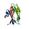
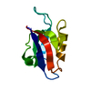
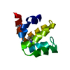
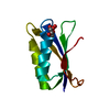
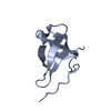
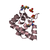
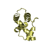
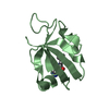
| Entry | Database: PDB / ID: 5d77 | ||||||
|---|---|---|---|---|---|---|---|
| Title | Structure of Mip6 RRM3 Domain | ||||||
 Components Components | RNA-binding protein MIP6 | ||||||
 Keywords Keywords | RNA BINDING PROTEIN / RNA Binding Domain | ||||||
| Function / homology |  Function and homology information Function and homology informationmRNA localization resulting in post-transcriptional regulation of gene expression / Deadenylation of mRNA / sporulation / prospore membrane / mRNA metabolic process / poly(A) binding / poly(U) RNA binding / Translation initiation complex formation / Nonsense Mediated Decay (NMD) independent of the Exon Junction Complex (EJC) / Nonsense Mediated Decay (NMD) enhanced by the Exon Junction Complex (EJC) ...mRNA localization resulting in post-transcriptional regulation of gene expression / Deadenylation of mRNA / sporulation / prospore membrane / mRNA metabolic process / poly(A) binding / poly(U) RNA binding / Translation initiation complex formation / Nonsense Mediated Decay (NMD) independent of the Exon Junction Complex (EJC) / Nonsense Mediated Decay (NMD) enhanced by the Exon Junction Complex (EJC) / L13a-mediated translational silencing of Ceruloplasmin expression / mRNA 3'-UTR binding / cytoplasmic stress granule / regulation of translation / ribonucleoprotein complex / RNA binding / nucleus / cytosol Similarity search - Function | ||||||
| Biological species |  | ||||||
| Method |  X-RAY DIFFRACTION / SYNCHROTRON / MOLECULAR REPLACEMENT / Resolution: 1.3 Å X-RAY DIFFRACTION / SYNCHROTRON / MOLECULAR REPLACEMENT / Resolution: 1.3 Å | ||||||
 Authors Authors | Mohamad, N. / Bravo, J. | ||||||
 Citation Citation | Journal: To Be Published Title: Structure of Mip6 RRM3 domain at 1.3 Authors: Mohamad, N. / Bravo, J. | ||||||
| History |
|
- Structure visualization
| Structure viewer | Molecule: MolmilJmol/JSmol |
|---|
- Downloads & links
-Download
| PDBx/mmCIF format | 5d77.cif.gz | 64.4 KB | Display | PDBx/mmCIF format |
|---|---|---|---|---|
| PDB format | pdb5d77.ent.gz | 46.8 KB | Display | PDB format |
| PDBx/mmJSON format | 5d77.json.gz | Tree view | PDBx/mmJSON format | |
| Others |  Other downloads Other downloads |
-Validation report
| Arichive directory | https://data.pdbj.org/pub/pdb/validation_reports/d7/5d77ftp://data.pdbj.org/pub/pdb/validation_reports/d7/5d77 | HTTPS FTP |
|---|
-Related structure data
| Related structure data |  3feyS S: Starting model for refinement |
|---|---|
| Similar structure data |


-Links
 PDBj
PDBj


- Assembly
| Deposited unit | 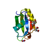
| ||||||||
|---|---|---|---|---|---|---|---|---|---|
| 1 |
| ||||||||
| Unit cell |
|
-Components
| #1: Protein | Mass: 9211.569 Da / Num. of mol.: 1 Source method: isolated from a genetically manipulated source Source: (gene. exp.) Gene: MIP6, YHR015W / Production host:  | ||||||
|---|---|---|---|---|---|---|---|
| #2: Chemical | ChemComp-CIT /   Mass: 192.124 Da / Num. of mol.: 1 / Source method: obtained synthetically / Formula: C6H8O7 Mass: 192.124 Da / Num. of mol.: 1 / Source method: obtained synthetically / Formula: C6H8O7 | ||||||
| #3: Chemical |   Mass: 62.005 Da / Num. of mol.: 2 / Source method: obtained synthetically / Formula: NO3 Mass: 62.005 Da / Num. of mol.: 2 / Source method: obtained synthetically / Formula: NO3#4: Chemical | ChemComp-NA / |   Mass: 22.990 Da / Num. of mol.: 1 / Source method: obtained synthetically / Formula: Na Mass: 22.990 Da / Num. of mol.: 1 / Source method: obtained synthetically / Formula: Na#5: Water | ChemComp-HOH / |  Mass: 18.015 Da / Num. of mol.: 51 / Source method: isolated from a natural source / Formula: H2O Mass: 18.015 Da / Num. of mol.: 51 / Source method: isolated from a natural source / Formula: H2OHas protein modification | Y | |
-Experimental details
-Experiment
| Experiment | Method: X-RAY DIFFRACTION |
|---|
- Sample preparation
| Crystal | Density Matthews: 1.64 Å3/Da / Density % sol: 25.12 % |
|---|---|
| Crystal grow | Temperature: 294 K / Method: vapor diffusion / pH: 6.5 Details: 1.6M Sodium Citrate Tribasic dihydrate pH 6.5; Ammonium Nitrate |
-Data collection
| Diffraction | Mean temperature: 100 K |
|---|---|
| Diffraction source | Source: SYNCHROTRON / Site: ALBA  / Beamline: XALOC / Wavelength: 0.97934 Å / Beamline: XALOC / Wavelength: 0.97934 Å |
| Detector | Type: DECTRIS PILATUS 6M / Detector: PIXEL / Date: Feb 20, 2015 |
| Radiation | Monochromator: Si(111) / Protocol: SINGLE WAVELENGTH / Monochromatic (M) / Laue (L): M / Scattering type: x-ray |
| Radiation wavelength | Wavelength: 0.97934 Å / Relative weight: 1 |
| Reflection | Resolution: 1.3→45.91 Å / Num. obs: 15814 / % possible obs: 99.4 % / Redundancy: 11.3 % / Rmerge(I) obs: 0.45 / Net I/σ(I): 25.1 |
| Reflection shell | Resolution: 1.3→1.35 Å / Redundancy: 5 % / Mean I/σ(I) obs: 3.5 / % possible all: 95.9 |
- Processing
| Software |
| |||||||||||||||||||||||||||||||||||||||||||||||||
|---|---|---|---|---|---|---|---|---|---|---|---|---|---|---|---|---|---|---|---|---|---|---|---|---|---|---|---|---|---|---|---|---|---|---|---|---|---|---|---|---|---|---|---|---|---|---|---|---|---|---|
| Refinement | Method to determine structure: MOLECULAR REPLACEMENT Starting model: 3FEY Resolution: 1.3→33.766 Å / SU ML: 0.12 / Cross valid method: FREE R-VALUE / σ(F): 1.35 / Phase error: 20.77 / Stereochemistry target values: ML
| |||||||||||||||||||||||||||||||||||||||||||||||||
| Solvent computation | Shrinkage radii: 0.9 Å / VDW probe radii: 1.11 Å / Solvent model: FLAT BULK SOLVENT MODEL | |||||||||||||||||||||||||||||||||||||||||||||||||
| Refinement step | Cycle: LAST / Resolution: 1.3→33.766 Å
| |||||||||||||||||||||||||||||||||||||||||||||||||
| Refine LS restraints |
| |||||||||||||||||||||||||||||||||||||||||||||||||
| LS refinement shell |
|
