 Movie
Movie Controller


+ Open data

- Basic information
| Entry | Database: PDB / ID: 6eto | ||||||
|---|---|---|---|---|---|---|---|
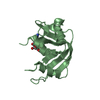
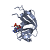
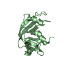
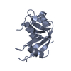
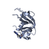
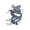
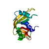
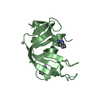
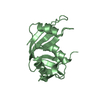
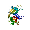
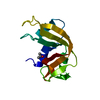
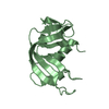
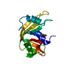
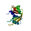
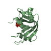
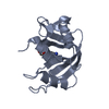
| Title | Atomic resolution structure of RNase A (data collection 5) | ||||||
 Components Components | Ribonuclease pancreatic | ||||||
 Keywords Keywords | HYDROLASE / Raman microspectroscopy / ribonuclease / atomic resolution / radiation damage / photodamage | ||||||
| Function / homology |  Function and homology information Function and homology informationpancreatic ribonuclease / ribonuclease A activity / RNA nuclease activity / nucleic acid binding / defense response to Gram-positive bacterium / lyase activity / extracellular region Similarity search - Function | ||||||
| Biological species |  | ||||||
| Method |  X-RAY DIFFRACTION / SYNCHROTRON / MOLECULAR REPLACEMENT / Resolution: 1.02 Å X-RAY DIFFRACTION / SYNCHROTRON / MOLECULAR REPLACEMENT / Resolution: 1.02 Å | ||||||
 Authors Authors | Caterino, M. / Vergara, A. / Merlino, A. | ||||||
 Citation Citation | Journal: Chembiochem / Year: 2018 Title: The Alkylquinolone Repertoire of Pseudomonas aeruginosa is Linked to Structural Flexibility of the FabH-like 2-Heptyl-3-hydroxy-4(1H)-quinolone (PQS) Biosynthesis Enzyme PqsBC. Authors: Witzgall, F. / Depke, T. / Hoffmann, M. / Empting, M. / Bronstrup, M. / Muller, R. / Blankenfeldt, W. | ||||||
| History |
|
- Structure visualization
| Structure viewer | Molecule: MolmilJmol/JSmol |
|---|
- Downloads & links
-Download
| PDBx/mmCIF format | 6eto.cif.gz | 100 KB | Display | PDBx/mmCIF format |
|---|---|---|---|---|
| PDB format | pdb6eto.ent.gz | 79 KB | Display | PDB format |
| PDBx/mmJSON format | 6eto.json.gz | Tree view | PDBx/mmJSON format | |
| Others |  Other downloads Other downloads |
-Validation report
| Arichive directory | https://data.pdbj.org/pub/pdb/validation_reports/et/6etoftp://data.pdbj.org/pub/pdb/validation_reports/et/6eto | HTTPS FTP |
|---|
-Related structure data
| Related structure data |  6eszC  6et0C  6et1C  6et2C  6et3C  1kf3S S: Starting model for refinement C: citing same article ( |
|---|---|
| Similar structure data |


-Links
 PDBj
PDBj


- Assembly
| Deposited unit | 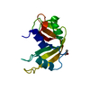
| ||||||||
|---|---|---|---|---|---|---|---|---|---|
| 1 |
| ||||||||
| Unit cell |
|
-Components
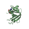
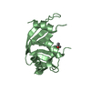
| #1: Protein | Mass: 13708.326 Da / Num. of mol.: 1 / Source method: isolated from a natural source / Source: (natural) | ||||
|---|---|---|---|---|---|
| #2: Chemical |   Mass: 60.095 Da / Num. of mol.: 3 / Source method: obtained synthetically / Formula: C3H8O / Comment: alkaloid*YM Mass: 60.095 Da / Num. of mol.: 3 / Source method: obtained synthetically / Formula: C3H8O / Comment: alkaloid*YM#3: Water | ChemComp-HOH / |  Mass: 18.015 Da / Num. of mol.: 211 / Source method: isolated from a natural source / Formula: H2O Mass: 18.015 Da / Num. of mol.: 211 / Source method: isolated from a natural source / Formula: H2OHas protein modification | Y | |
-Experimental details
-Experiment
| Experiment | Method: X-RAY DIFFRACTION / Number of used crystals: 1 |
|---|
- Sample preparation
| Crystal | Density Matthews: 2.04 Å3/Da / Density % sol: 39.68 % |
|---|---|
| Crystal grow | Temperature: 293 K / Method: liquid diffusion Details: Free-interface diffusion of a 10 uL of 30 mg ml-1 protein solution, 20 mM sodium citrate at pH 5.3 agaist 10 ul isopropanol 99.9% isopropanol. |
-Data collection
| Diffraction | Mean temperature: 100 K |
|---|---|
| Diffraction source | Source: SYNCHROTRON / Site: SLS  / Beamline: X10SA / Wavelength: 0.7749 Å / Beamline: X10SA / Wavelength: 0.7749 Å |
| Detector | Type: DECTRIS PILATUS3 6M / Detector: PIXEL / Date: Sep 26, 2011 |
| Radiation | Protocol: SINGLE WAVELENGTH / Monochromatic (M) / Laue (L): M / Scattering type: x-ray |
| Radiation wavelength | Wavelength: 0.7749 Å / Relative weight: 1 |
| Reflection | Resolution: 1.02→50 Å / Num. obs: 54830 / % possible obs: 98.9 % / Redundancy: 6.6 % / Rmerge(I) obs: 0.094 / Net I/σ(I): 10.15 |
| Reflection shell | Resolution: 1.02→1.06 Å / Redundancy: 6.4 % / Rmerge(I) obs: 0.451 / Mean I/σ(I) obs: 4.29 / % possible all: 97.7 |
- Processing
| Software |
| ||||||||||||||||
|---|---|---|---|---|---|---|---|---|---|---|---|---|---|---|---|---|---|
| Refinement | Method to determine structure: MOLECULAR REPLACEMENT Starting model: 1KF3 Resolution: 1.02→50 Å / Cross valid method: FREE R-VALUE
| ||||||||||||||||
| Displacement parameters | Biso mean: 19.17 Å2 | ||||||||||||||||
| Refinement step | Cycle: LAST / Resolution: 1.02→50 Å
|
