 Movie
Movie Controller


+ Open data

- Basic information
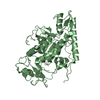
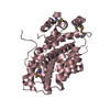
| Entry | Database: PDB / ID: 6en7 | ||||||
|---|---|---|---|---|---|---|---|
| Title | Crystal structure of the ribosome assembly factor Nsa1 | ||||||
 Components Components | Ribosome biogenesis protein NSA1 | ||||||
 Keywords Keywords | CHAPERONE / Ribosome / biogenesis | ||||||
| Function / homology | WDR74/Nsa1 / preribosome, large subunit precursor / ribosomal large subunit biogenesis / rRNA processing / WD40-repeat-containing domain superfamily / nucleolus / nucleus / Ribosome biogenesis protein NSA1 Function and homology information Function and homology information | ||||||
| Biological species |  | ||||||
| Method |  X-RAY DIFFRACTION / SYNCHROTRON / MOLECULAR REPLACEMENT / Resolution: 2.403 Å X-RAY DIFFRACTION / SYNCHROTRON / MOLECULAR REPLACEMENT / Resolution: 2.403 Å | ||||||
 Authors Authors | Altegoer, F. / Bange, G. | ||||||
 Citation Citation | Journal: Cell / Year: 2017 Title: Visualizing the Assembly Pathway of Nucleolar Pre-60S Ribosomes. Authors: Lukas Kater / Matthias Thoms / Clara Barrio-Garcia / Jingdong Cheng / Sherif Ismail / Yasar Luqman Ahmed / Gert Bange / Dieter Kressler / Otto Berninghausen / Irmgard Sinning / Ed Hurt / Roland Beckmann /  Abstract: Eukaryotic 60S ribosomal subunits are comprised of three rRNAs and ∼50 ribosomal proteins. The initial steps of their formation take place in the nucleolus, but, owing to a lack of structural ...Eukaryotic 60S ribosomal subunits are comprised of three rRNAs and ∼50 ribosomal proteins. The initial steps of their formation take place in the nucleolus, but, owing to a lack of structural information, this process is poorly understood. Using cryo-EM, we solved structures of early 60S biogenesis intermediates at 3.3 Å to 4.5 Å resolution, thereby providing insights into their sequential folding and assembly pathway. Besides revealing distinct immature rRNA conformations, we map 25 assembly factors in six different assembly states. Notably, the Nsa1-Rrp1-Rpf1-Mak16 module stabilizes the solvent side of the 60S subunit, and the Erb1-Ytm1-Nop7 complex organizes and connects through Erb1's meandering N-terminal extension, eight assembly factors, three ribosomal proteins, and three 25S rRNA domains. Our structural snapshots reveal the order of integration and compaction of the six major 60S domains within early nucleolar 60S particles developing stepwise from the solvent side around the exit tunnel to the central protuberance. | ||||||
| History |
|
- Structure visualization
| Structure viewer | Molecule: MolmilJmol/JSmol |
|---|
- Downloads & links
-Download
| PDBx/mmCIF format | 6en7.cif.gz | 93.3 KB | Display | PDBx/mmCIF format |
|---|---|---|---|---|
| PDB format | pdb6en7.ent.gz | 68.7 KB | Display | PDB format |
| PDBx/mmJSON format | 6en7.json.gz | Tree view | PDBx/mmJSON format | |
| Others |  Other downloads Other downloads |
-Validation report
| Arichive directory | https://data.pdbj.org/pub/pdb/validation_reports/en/6en7ftp://data.pdbj.org/pub/pdb/validation_reports/en/6en7 | HTTPS FTP |
|---|
-Related structure data
| Related structure data | 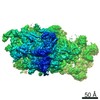 3888C 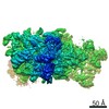 3889C 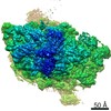 3890C 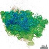 3891C  3892C  3893C  6elzC  6em1C  6em3C  6em4C  6em5C  6emfC 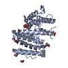 6emgC  5sumS S: Starting model for refinement C: citing same article ( |
|---|---|
| Similar structure data |

-Links
 PDBj
PDBj

- Assembly
| Deposited unit | 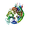
| ||||||||
|---|---|---|---|---|---|---|---|---|---|
| 1 |
| ||||||||
| Unit cell |
|
-Components
| #1: Protein | Mass: 51982.457 Da / Num. of mol.: 1 Source method: isolated from a genetically manipulated source Source: (gene. exp.) Gene: NSA1, YGL111W, G2990 / Production host:  |
|---|---|
| #2: Water | ChemComp-HOH /  Mass: 18.015 Da / Num. of mol.: 230 / Source method: isolated from a natural source / Formula: H2O Mass: 18.015 Da / Num. of mol.: 230 / Source method: isolated from a natural source / Formula: H2O |
-Experimental details
-Experiment
| Experiment | Method: X-RAY DIFFRACTION / Number of used crystals: 1 |
|---|
- Sample preparation
| Crystal | Density Matthews: 2.31 Å3/Da / Density % sol: 46.83 % |
|---|---|
| Crystal grow | Temperature: 294 K / Method: vapor diffusion, sitting drop / Details: 0.1 M MES pH 6.5, 25% w/v PEG-1000 |
-Data collection
| Diffraction | Mean temperature: 100 K |
|---|---|
| Diffraction source | Source: SYNCHROTRON / Site: ESRF  / Beamline: ID29 / Wavelength: 0.987 Å / Beamline: ID29 / Wavelength: 0.987 Å |
| Detector | Type: DECTRIS PILATUS3 6M / Detector: PIXEL / Date: May 19, 2016 |
| Radiation | Protocol: SINGLE WAVELENGTH / Monochromatic (M) / Laue (L): M / Scattering type: x-ray |
| Radiation wavelength | Wavelength: 0.987 Å / Relative weight: 1 |
| Reflection | Resolution: 2.4→59.5 Å / Num. obs: 15750 / % possible obs: 99.9 % / Redundancy: 7.1 % / CC1/2: 0.999 / Rmerge(I) obs: 0.04384 / Net I/σ(I): 34.78 |
| Reflection shell | Resolution: 2.4→2.489 Å / Redundancy: 5.6 % / Rmerge(I) obs: 0.1126 / Mean I/σ(I) obs: 14.02 / Num. unique obs: 1543 / CC1/2: 0.992 / % possible all: 99.23 |
- Processing
| Software |
| ||||||||||||||||||||||||||||||||||||||||||
|---|---|---|---|---|---|---|---|---|---|---|---|---|---|---|---|---|---|---|---|---|---|---|---|---|---|---|---|---|---|---|---|---|---|---|---|---|---|---|---|---|---|---|---|
| Refinement | Method to determine structure: MOLECULAR REPLACEMENT Starting model: 5SUM Resolution: 2.403→46.408 Å / SU ML: 0.21 / Cross valid method: FREE R-VALUE / σ(F): 1.26 / Phase error: 19.4
| ||||||||||||||||||||||||||||||||||||||||||
| Solvent computation | Shrinkage radii: 0.9 Å / VDW probe radii: 1.11 Å | ||||||||||||||||||||||||||||||||||||||||||
| Refinement step | Cycle: LAST / Resolution: 2.403→46.408 Å
| ||||||||||||||||||||||||||||||||||||||||||
| Refine LS restraints |
| ||||||||||||||||||||||||||||||||||||||||||
| LS refinement shell |
|
