[English] 日本語
Yorodumi
- PDB-6eho: Dimer of the Sortilin Vps10p domain at low pH -

+
Open data


ID or keywords:

Loading...

-
Basic information

Entry
Database: PDB / ID: 6eho
TitleDimer of the Sortilin Vps10p domain at low pH
ComponentsSortilin
KeywordsPROTEIN BINDING / PROTEIN SORTING RECEPTOR / 10-bladed beta-propeller / Vps10p-D / Endocytosis / Endosome / Glycoprotein / Golgi apparatus / Lysosome / Membrane / Receptor / Transmembrane / SIGNALING PROTEIN
Function / homology
Function and homology information


neurotensin receptor activity, non-G protein-coupled / negative regulation of lipoprotein lipase activity / myotube differentiation / cerebellar climbing fiber to Purkinje cell synapse / plasma membrane to endosome transport / retromer complex binding / maintenance of synapse structure / Golgi to endosome transport / nerve growth factor receptor activity / Golgi to lysosome transport ...neurotensin receptor activity, non-G protein-coupled / negative regulation of lipoprotein lipase activity / myotube differentiation / cerebellar climbing fiber to Purkinje cell synapse / plasma membrane to endosome transport / retromer complex binding / maintenance of synapse structure / Golgi to endosome transport / nerve growth factor receptor activity / Golgi to lysosome transport / vesicle organization / endosome transport via multivesicular body sorting pathway / nerve growth factor binding / protein targeting to lysosome / trans-Golgi network transport vesicle / clathrin-coated vesicle / negative regulation of fat cell differentiation / Golgi cisterna membrane / Golgi Associated Vesicle Biogenesis / endosome to lysosome transport / glucose import / neurotrophin TRK receptor signaling pathway / extrinsic apoptotic signaling pathway via death domain receptors / neuropeptide signaling pathway / clathrin-coated pit / ossification / response to insulin / endocytosis / cytoplasmic vesicle / regulation of gene expression / nuclear membrane / lysosome / early endosome / endosome membrane / G protein-coupled receptor signaling pathway / lysosomal membrane / endoplasmic reticulum membrane / perinuclear region of cytoplasm / Golgi apparatus / enzyme binding / cell surface / membrane / plasma membrane / cytosol
Similarity search - Function
VPS10 / Sortilin, C-terminal / Sortilin, N-terminal / Sortilin, neurotensin receptor 3, C-terminal / Sortilin, neurotensin receptor 3, / VPS10 / WD40/YVTN repeat-like-containing domain superfamily
Similarity search - Domain/homology
Biological speciesHomo sapiens (human)
MethodX-RAY DIFFRACTION / SYNCHROTRON / MOLECULAR REPLACEMENT / Resolution: 3.5 Å
AuthorsThirup, S.S. / Quistgaard, E.H. / Januliene, D. / Andersen, J.L. / Nielsen, J.A.
Funding support Denmark, 1items
OrganizationGrant numberCountry
Lundbeck Foundation Denmark
Citation
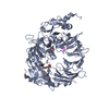
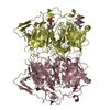
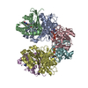
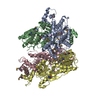
Journal: Structure / Year: 2017
Title: Acidic Environment Induces Dimerization and Ligand Binding Site Collapse in the Vps10p Domain of Sortilin.
Authors: Dovile Januliene / Jacob Lauwring Andersen / Jeppe Achton Nielsen / Esben Meldgaard Quistgaard / Maria Hansen / Dorthe Strandbygaard / Arne Moeller / Claus Munck Petersen / Peder Madsen / Søren Skou Thirup /
Abstract: Sortilin is a neuronal receptor involved in transmembrane signaling, endocytosis, and intracellular sorting of proteins. It cycles through a number of cellular compartments where it encounters ...Sortilin is a neuronal receptor involved in transmembrane signaling, endocytosis, and intracellular sorting of proteins. It cycles through a number of cellular compartments where it encounters various acidic conditions. The crystal structure of the sortilin ectodomain has previously been determined at neutral pH. Here, we present the 3.5-Å resolution crystal structure of sortilin at pH 5.5, which represents an environment similar to that of late endosomes, where ligands are released. The structure reveals an overall distortion of the 10-bladed β-propeller domain. This distortion and specific conformational changes, caused by protonation of a number of histidine residues, render the currently known binding sites unavailable for ligand binding. Access to the binding sites is furthermore blocked by a reversible and pH-dependent formation of tight sortilin dimers, also confirmed by electron microscopy, size-exclusion chromatography, and mutational studies. This study reveals how sortilin binding sites are disrupted and explains pH-dependent ligand affinity.
History
DepositionSep 14, 2017Deposition site: PDBE / Processing site: PDBE
Revision 1.0Dec 6, 2017Provider: repository / Type: Initial release
Revision 1.1Dec 13, 2017Group: Database references / Category: citation
Item: _citation.journal_volume / _citation.page_first / _citation.page_last
Revision 2.0Jul 29, 2020Group: Atomic model / Data collection ...Atomic model / Data collection / Derived calculations / Structure summary
Category: atom_site / chem_comp ...atom_site / chem_comp / entity / pdbx_branch_scheme / pdbx_chem_comp_identifier / pdbx_entity_branch / pdbx_entity_branch_descriptor / pdbx_entity_branch_link / pdbx_entity_branch_list / pdbx_entity_nonpoly / pdbx_nonpoly_scheme / pdbx_struct_assembly_gen / struct_asym / struct_conn / struct_site / struct_site_gen
Item: _atom_site.B_iso_or_equiv / _atom_site.Cartn_x ..._atom_site.B_iso_or_equiv / _atom_site.Cartn_x / _atom_site.Cartn_y / _atom_site.Cartn_z / _atom_site.auth_asym_id / _atom_site.auth_seq_id / _atom_site.label_asym_id / _atom_site.label_entity_id / _chem_comp.name / _chem_comp.type / _entity.formula_weight / _entity.pdbx_description / _entity.pdbx_number_of_molecules / _entity.type / _pdbx_struct_assembly_gen.asym_id_list / _struct_conn.pdbx_role / _struct_conn.ptnr1_auth_asym_id / _struct_conn.ptnr1_auth_seq_id / _struct_conn.ptnr1_label_asym_id / _struct_conn.ptnr2_auth_asym_id / _struct_conn.ptnr2_auth_seq_id / _struct_conn.ptnr2_label_asym_id
Description: Carbohydrate remediation / Provider: repository / Type: Remediation
Revision 2.1Jan 17, 2024Group: Data collection / Database references ...Data collection / Database references / Derived calculations / Refinement description / Structure summary
Category: chem_comp / chem_comp_atom ...chem_comp / chem_comp_atom / chem_comp_bond / database_2 / pdbx_initial_refinement_model / struct_conn
Item: _chem_comp.pdbx_synonyms / _database_2.pdbx_DOI ..._chem_comp.pdbx_synonyms / _database_2.pdbx_DOI / _database_2.pdbx_database_accession / _struct_conn.pdbx_leaving_atom_flag

-
Structure visualization

Structure viewerMolecule:
MolmilJmol/JSmol

Downloads & links

-
Assembly

Deposited unit
A: Sortilin
hetero molecules


Theoretical massNumber of molelcules
Total (without water)82,3034
Polymers80,3811
Non-polymers1,9223
Water0
1
A: Sortilin
hetero molecules

A: Sortilin
hetero molecules


Theoretical massNumber of molelcules
Total (without water)164,6058
Polymers160,7622
Non-polymers3,8446
Water0
TypeNameSymmetry operationNumber
identity operation1_555x,y,z1
crystal symmetry operation16_555x,-y,-z+1/21
Buried area9230 Å2
ΔGint33 kcal/mol
Surface area56890 Å2
MethodPISA
Unit cell
Length a, b, c (Å)189.010, 189.010, 189.010
Angle α, β, γ (deg.)90.00, 90.00, 90.00
Int Tables number199
Space group name H-MI213

-
Components

#1: Protein Sortilin / 100 kDa NT receptor / Glycoprotein 95 / Gp95 / Neurotensin receptor 3 / NTR3


Mass: 80380.797 Da / Num. of mol.: 1 / Mutation: R43G , R44G
Source method: isolated from a genetically manipulated source
Source: (gene. exp.) Homo sapiens (human) / Gene: SORT1 / Production host: 
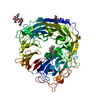
Cricetulus griseus (Chinese hamster) / References: UniProt: Q99523
#2: Polysaccharide alpha-D-mannopyranose-(1-3)-[alpha-D-mannopyranose-(1-6)]beta-D-mannopyranose-(1-4)-2-acetamido-2- ...alpha-D-mannopyranose-(1-3)-[alpha-D-mannopyranose-(1-6)]beta-D-mannopyranose-(1-4)-2-acetamido-2-deoxy-beta-D-glucopyranose-(1-4)-2-acetamido-2-deoxy-beta-D-glucopyranose


Type: oligosaccharide / Mass: 910.823 Da / Num. of mol.: 1
Source method: isolated from a genetically manipulated source
DescriptorTypeProgram
DManpa1-3[DManpa1-6]DManpb1-4DGlcpNAcb1-4DGlcpNAcb1-Glycam Condensed SequenceGMML 1.0
WURCS=2.0/3,5,4/[a2122h-1b_1-5_2*NCC/3=O][a1122h-1b_1-5][a1122h-1a_1-5]/1-1-2-3-3/a4-b1_b4-c1_c3-d1_c6-e1WURCSPDB2Glycan 1.1.0
[]{[(4+1)][b-D-GlcpNAc]{[(4+1)][b-D-GlcpNAc]{[(4+1)][b-D-Manp]{[(3+1)][a-D-Manp]{}[(6+1)][a-D-Manp]{}}}}}LINUCSPDB-CARE
#3: Polysaccharide beta-D-mannopyranose-(1-4)-2-acetamido-2-deoxy-beta-D-glucopyranose-(1-4)-2-acetamido-2-deoxy-beta- ...beta-D-mannopyranose-(1-4)-2-acetamido-2-deoxy-beta-D-glucopyranose-(1-4)-2-acetamido-2-deoxy-beta-D-glucopyranose


Type: oligosaccharide / Mass: 586.542 Da / Num. of mol.: 1
Source method: isolated from a genetically manipulated source
DescriptorTypeProgram
DManpb1-4DGlcpNAcb1-4DGlcpNAcb1-Glycam Condensed SequenceGMML 1.0
WURCS=2.0/2,3,2/[a2122h-1b_1-5_2*NCC/3=O][a1122h-1b_1-5]/1-1-2/a4-b1_b4-c1WURCSPDB2Glycan 1.1.0
[]{[(4+1)][b-D-GlcpNAc]{[(4+1)][b-D-GlcpNAc]{[(4+1)][b-D-Manp]{}}}}LINUCSPDB-CARE
#4: Polysaccharide 2-acetamido-2-deoxy-beta-D-glucopyranose-(1-4)-2-acetamido-2-deoxy-beta-D-glucopyranose


Type: oligosaccharide / Mass: 424.401 Da / Num. of mol.: 1
Source method: isolated from a genetically manipulated source
DescriptorTypeProgram
DGlcpNAcb1-4DGlcpNAcb1-Glycam Condensed SequenceGMML 1.0
WURCS=2.0/1,2,1/[a2122h-1b_1-5_2*NCC/3=O]/1-1/a4-b1WURCSPDB2Glycan 1.1.0
[]{[(4+1)][b-D-GlcpNAc]{[(4+1)][b-D-GlcpNAc]{}}}LINUCSPDB-CARE

-
Experimental details

-
Experiment

ExperimentMethod: X-RAY DIFFRACTION / Number of used crystals: 1

-
Sample preparation

CrystalDensity Matthews: 3.5 Å3/Da / Density % sol: 64.86 %
Crystal growTemperature: 291 K / Method: vapor diffusion, sitting drop / pH: 5.5 / Details: 2.0 Ammonium sulphate / PH range: 5.0 - 5.5

-
Data collection

DiffractionMean temperature: 100 K
Diffraction sourceSource: SYNCHROTRON / Site: SLS / Beamline: X06SA / Wavelength: 1 Å
DetectorType: PSI PILATUS 6M / Detector: PIXEL / Date: Dec 5, 2008
RadiationProtocol: SINGLE WAVELENGTH / Monochromatic (M) / Laue (L): M / Scattering type: x-ray
Radiation wavelengthWavelength: 1 Å / Relative weight: 1
ReflectionResolution: 3.5→94.505 Å / Num. obs: 14135 / % possible obs: 98.6 % / Redundancy: 3.9 % / Rmerge(I) obs: 0.052 / Net I/σ(I): 17.4
Reflection shellResolution: 3.5→3.62 Å / Rmerge(I) obs: 0.59

-
Processing

Software
NameVersionClassification
PHENIX(dev_2614: ???)refinement
XDSdata reduction
XSCALEdata scaling
PHASERphasing
RefinementMethod to determine structure: MOLECULAR REPLACEMENT
Starting model: 3f6k
Resolution: 3.5→94.505 Å / SU ML: 0.44 / Cross valid method: FREE R-VALUE / σ(F): 2 / Phase error: 30.77 / Stereochemistry target values: ML
RfactorNum. reflection% reflection
Rfree0.272 566 4.01 %
Rwork0.2315 --
obs0.2333 14129 98.56 %
Solvent computationShrinkage radii: 0.9 Å / VDW probe radii: 1.11 Å / Solvent model: FLAT BULK SOLVENT MODEL
Refinement stepCycle: LAST / Resolution: 3.5→94.505 Å
ProteinNucleic acidLigandSolventTotal
Num. atoms5035 0 128 0 5163
Refine LS restraints
Refine-IDTypeDev idealNumber
X-RAY DIFFRACTIONf_bond_d0.0025301
X-RAY DIFFRACTIONf_angle_d0.5537208
X-RAY DIFFRACTIONf_dihedral_angle_d10.5063110
X-RAY DIFFRACTIONf_chiral_restr0.044811
X-RAY DIFFRACTIONf_plane_restr0.003911
LS refinement shell
Resolution (Å)Rfactor RfreeNum. reflection RfreeRfactor RworkNum. reflection RworkRefine-ID% reflection obs (%)
3.5002-3.85240.28321410.24853369X-RAY DIFFRACTION99
3.8524-4.40990.29581400.233381X-RAY DIFFRACTION99
4.4099-5.55590.22651420.22933390X-RAY DIFFRACTION99
5.5559-94.53940.28591430.22993423X-RAY DIFFRACTION97
Refinement TLS params.

Method: refined / Refine-ID: X-RAY DIFFRACTION

IDL112)L122)L132)L222)L232)L332)S11 (Å °)S12 (Å °)S13 (Å °)S21 (Å °)S22 (Å °)S23 (Å °)S31 (Å °)S32 (Å °)S33 (Å °)T112)T122)T132)T222)T232)T332)Origin x (Å)Origin y (Å)Origin z (Å)
12.29020.07430.39575.1036-0.99182.61960.0739-0.25560.2224-0.0223-0.11640.18050.0473-0.1554-0.01010.7365-0.08640.10590.94740.14340.986848.872417.406732.3152
22.19810.8541-0.47494.02580.8261.2233-0.0894-0.41530.4017-0.0520.1062-1.0111-0.46550.32090.24311.0617-0.30530.01221.35990.26441.57779.615130.153328.9132
33.2544-1.1779-1.39794.76422.071.73260.1390.6423-0.9511-0.5758-0.34890.50380.6191-0.07570.09741.2458-0.24240.14911.08180.16961.066744.8715-4.411626.4959
43.4770.73510.95752.06860.2272-0.13680.04640.6627-0.0724-0.6924-0.332-0.83680.44640.76510.33921.29960.26780.41921.3590.31461.303574.0228-5.720624.7274
54.6273-1.0235-0.60082.9267-0.38863.8340.1772-0.56530.19450.5416-0.3683-1.0382-0.46890.82640.14730.9718-0.27830.02391.18840.18021.354471.20922.512840.1035
Refinement TLS group
IDRefine-IDRefine TLS-IDSelection details
1X-RAY DIFFRACTION1chain 'A' and (resid 422 through 617 )
2X-RAY DIFFRACTION2chain 'A' and (resid 618 through 709 )
3X-RAY DIFFRACTION3chain 'A' and (resid 52 through 129 )
4X-RAY DIFFRACTION4chain 'A' and (resid 130 through 307 )
5X-RAY DIFFRACTION5chain 'A' and (resid 308 through 421 )

+
About Yorodumi

-
News

-
Feb 9, 2022. New format data for meta-information of EMDB entries

New format data for meta-information of EMDB entries

  • Version 3 of the EMDB header file is now the official format.
  • The previous official version 1.9 will be removed from the archive.

Related info.:EMDB header

External links:wwPDB to switch to version 3 of the EMDB data model

-
Aug 12, 2020. Covid-19 info

Covid-19 info

URL: https://pdbj.org/emnavi/covid19.php

New page: Covid-19 featured information page in EM Navigator.

Related info.:Covid-19 info / Mar 5, 2020. Novel coronavirus structure data

+
Mar 5, 2020. Novel coronavirus structure data

Novel coronavirus structure data

Related info.:Yorodumi Speices / Aug 12, 2020. Covid-19 info

External links:COVID-19 featured content - PDBj / Molecule of the Month (242):Coronavirus Proteases

+
Jan 31, 2019. EMDB accession codes are about to change! (news from PDBe EMDB page)

EMDB accession codes are about to change! (news from PDBe EMDB page)

  • The allocation of 4 digits for EMDB accession codes will soon come to an end. Whilst these codes will remain in use, new EMDB accession codes will include an additional digit and will expand incrementally as the available range of codes is exhausted. The current 4-digit format prefixed with “EMD-” (i.e. EMD-XXXX) will advance to a 5-digit format (i.e. EMD-XXXXX), and so on. It is currently estimated that the 4-digit codes will be depleted around Spring 2019, at which point the 5-digit format will come into force.
  • The EM Navigator/Yorodumi systems omit the EMD- prefix.

Related info.:Q: What is EMD? / ID/Accession-code notation in Yorodumi/EM Navigator

External links:EMDB Accession Codes are Changing Soon! / Contact to PDBj

+
Jul 12, 2017. Major update of PDB

Major update of PDB

  • wwPDB released updated PDB data conforming to the new PDBx/mmCIF dictionary.
  • This is a major update changing the version number from 4 to 5, and with Remediation, in which all the entries are updated.
  • In this update, many items about electron microscopy experimental information are reorganized (e.g. em_software).
  • Now, EM Navigator and Yorodumi are based on the updated data.

External links:wwPDB Remediation / Enriched Model Files Conforming to OneDep Data Standards Now Available in the PDB FTP Archive

-
Yorodumi

Thousand views of thousand structures

  • Yorodumi is a browser for structure data from EMDB, PDB, SASBDB, etc.
  • This page is also the successor to EM Navigator detail page, and also detail information page/front-end page for Omokage search.
  • The word "yorodu" (or yorozu) is an old Japanese word meaning "ten thousand". "mi" (miru) is to see.

Related info.:EMDB / PDB / SASBDB / Comparison of 3 databanks / Yorodumi Search / Aug 31, 2016. New EM Navigator & Yorodumi / Yorodumi Papers / Jmol/JSmol / Function and homology information / Changes in new EM Navigator and Yorodumi

Read more