 Movie
Movie Controller

[English] 日本語
 Yorodumi
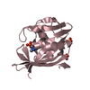
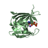
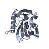
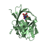
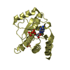
- PDB-6ehh: Crystal structure of mouse MTH1 mutant L116M with inhibitor TH588 -
+ Open data

- Basic information
| Entry | Database: PDB / ID: 6ehh | |||||||||||||||||||||||||||||||||
|---|---|---|---|---|---|---|---|---|---|---|---|---|---|---|---|---|---|---|---|---|---|---|---|---|---|---|---|---|---|---|---|---|---|---|
| Title | Crystal structure of mouse MTH1 mutant L116M with inhibitor TH588 | |||||||||||||||||||||||||||||||||
 Components Components | 7,8-dihydro-8-oxoguanine triphosphatase | |||||||||||||||||||||||||||||||||
 Keywords Keywords | HYDROLASE / Inhibitor / mutant / MTH1 / mouse | |||||||||||||||||||||||||||||||||
| Function / homology |  Function and homology information Function and homology informationPhosphate bond hydrolysis by NUDT proteins / 2-hydroxy-ATP hydrolase activity / 2-hydroxy-dATP hydrolase activity / N6-methyl-(d)ATP hydrolase activity / O6-methyl-dGTP hydrolase activity / 2-hydroxy-dATP diphosphatase / dATP diphosphatase activity / ATP diphosphatase activity / 8-oxo-7,8-dihydrodeoxyguanosine triphosphate pyrophosphatase activity / 8-oxo-7,8-dihydroguanosine triphosphate pyrophosphatase activity ...Phosphate bond hydrolysis by NUDT proteins / 2-hydroxy-ATP hydrolase activity / 2-hydroxy-dATP hydrolase activity / N6-methyl-(d)ATP hydrolase activity / O6-methyl-dGTP hydrolase activity / 2-hydroxy-dATP diphosphatase / dATP diphosphatase activity / ATP diphosphatase activity / 8-oxo-7,8-dihydrodeoxyguanosine triphosphate pyrophosphatase activity / 8-oxo-7,8-dihydroguanosine triphosphate pyrophosphatase activity / DNA protection / hydrolase activity, acting on acid anhydrides, in phosphorus-containing anhydrides / purine nucleoside catabolic process / snoRNA binding / Hydrolases; Acting on acid anhydrides; In phosphorus-containing anhydrides / acrosomal vesicle / nuclear membrane / mitochondrial matrix / mitochondrion / : / metal ion binding / nucleus / cytosol / cytoplasm Similarity search - Function | |||||||||||||||||||||||||||||||||
| Biological species |  | |||||||||||||||||||||||||||||||||
| Method |  X-RAY DIFFRACTION / SYNCHROTRON / MOLECULAR REPLACEMENT / Resolution: 2.4 Å X-RAY DIFFRACTION / SYNCHROTRON / MOLECULAR REPLACEMENT / Resolution: 2.4 Å | |||||||||||||||||||||||||||||||||
 Authors Authors | Gustafsson, R. / Narwal, M. / Jemth, A.-S. / Almlof, I. / Warpman Berglund, U. / Helleday, T. / Stenmark, P. | |||||||||||||||||||||||||||||||||
| Funding support |  Sweden, 10items Sweden, 10items
| |||||||||||||||||||||||||||||||||
 Citation Citation | Journal: Biochemistry / Year: 2018 Title: Crystal Structures and Inhibitor Interactions of Mouse and Dog MTH1 Reveal Species-Specific Differences in Affinity. Authors: Narwal, M. / Jemth, A.S. / Gustafsson, R. / Almlof, I. / Warpman Berglund, U. / Helleday, T. / Stenmark, P. | |||||||||||||||||||||||||||||||||
| History |
|
- Structure visualization
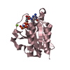
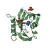
| Structure viewer | Molecule: MolmilJmol/JSmol |
|---|
- Downloads & links
-Download
| PDBx/mmCIF format | 6ehh.cif.gz | 148.7 KB | Display | PDBx/mmCIF format |
|---|---|---|---|---|
| PDB format | pdb6ehh.ent.gz | 115.4 KB | Display | PDB format |
| PDBx/mmJSON format | 6ehh.json.gz | Tree view | PDBx/mmJSON format | |
| Others |  Other downloads Other downloads |
-Validation report
| Arichive directory | https://data.pdbj.org/pub/pdb/validation_reports/eh/6ehhftp://data.pdbj.org/pub/pdb/validation_reports/eh/6ehh | HTTPS FTP |
|---|
-Related structure data
| Related structure data |  5mzeC  5mzfC  5mzgSC C: citing same article ( S: Starting model for refinement |
|---|---|
| Similar structure data |


-Links
 PDBj
PDBj

- Assembly
| Deposited unit | 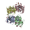
| ||||||||
|---|---|---|---|---|---|---|---|---|---|
| 1 | 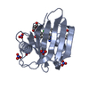
| ||||||||
| 2 | 
| ||||||||
| 3 | 
| ||||||||
| 4 | 
| ||||||||
| Unit cell |
|
-Components
-Protein , 1 types, 4 molecules ABCD
| #1: Protein | Mass: 20119.699 Da / Num. of mol.: 4 / Mutation: L116M Source method: isolated from a genetically manipulated source Source: (gene. exp.)  References: UniProt: P53368, 8-oxo-dGTP diphosphatase, 2-hydroxy-dATP diphosphatase |
|---|
-Non-polymers , 7 types, 298 molecules 


| #2: Chemical | ChemComp-2GE /  Mass: 295.167 Da / Num. of mol.: 4 / Source method: obtained synthetically / Formula: C13H12Cl2N4 / Feature type: SUBJECT OF INVESTIGATION Mass: 295.167 Da / Num. of mol.: 4 / Source method: obtained synthetically / Formula: C13H12Cl2N4 / Feature type: SUBJECT OF INVESTIGATION#3: Chemical | ChemComp-NO3 /  Mass: 62.005 Da / Num. of mol.: 19 / Source method: obtained synthetically / Formula: NO3 Mass: 62.005 Da / Num. of mol.: 19 / Source method: obtained synthetically / Formula: NO3#4: Chemical | ChemComp-PEG /  Mass: 106.120 Da / Num. of mol.: 5 / Source method: obtained synthetically / Formula: C4H10O3 Mass: 106.120 Da / Num. of mol.: 5 / Source method: obtained synthetically / Formula: C4H10O3#5: Chemical | ChemComp-GOL /  Mass: 92.094 Da / Num. of mol.: 4 / Source method: obtained synthetically / Formula: C3H8O3 Mass: 92.094 Da / Num. of mol.: 4 / Source method: obtained synthetically / Formula: C3H8O3#6: Chemical | ChemComp-CU /  Mass: 63.546 Da / Num. of mol.: 4 / Source method: obtained synthetically / Formula: Cu Mass: 63.546 Da / Num. of mol.: 4 / Source method: obtained synthetically / Formula: Cu#7: Chemical | ChemComp-MG / |  Mass: 24.305 Da / Num. of mol.: 1 / Source method: obtained synthetically / Formula: Mg Mass: 24.305 Da / Num. of mol.: 1 / Source method: obtained synthetically / Formula: Mg#8: Water | ChemComp-HOH / | Mass: 18.015 Da / Num. of mol.: 261 / Source method: isolated from a natural source / Formula: H2O |
|---|
-Experimental details
-Experiment
| Experiment | Method: X-RAY DIFFRACTION / Number of used crystals: 1 |
|---|
- Sample preparation
| Crystal | Density Matthews: 2.9 Å3/Da / Density % sol: 57.61 % |
|---|---|
| Crystal grow | Temperature: 293 K / Method: vapor diffusion, sitting drop Details: 0.2 M Ammonium nitrate, 34% (w/v) PEG 3350, 0.01 M copper (II) chloride dihydrate, 2 mM TCEP, 10 mM TH588, 6 mM MgCL2. |
-Data collection
| Diffraction | Mean temperature: 100 K |
|---|---|
| Diffraction source | Source: SYNCHROTRON / Site: BESSY  / Beamline: 14.1 / Wavelength: 0.9184 Å / Beamline: 14.1 / Wavelength: 0.9184 Å |
| Detector | Type: DECTRIS PILATUS 6M / Detector: PIXEL / Date: Aug 15, 2017 |
| Radiation | Protocol: SINGLE WAVELENGTH / Monochromatic (M) / Laue (L): M / Scattering type: x-ray |
| Radiation wavelength | Wavelength: 0.9184 Å / Relative weight: 1 |
| Reflection | Resolution: 2.4→47.4 Å / Num. obs: 35400 / % possible obs: 99 % / Redundancy: 3.9 % / CC1/2: 0.981 / Rrim(I) all: 0.271 / Net I/σ(I): 5 |
| Reflection shell | Resolution: 2.4→2.49 Å / Redundancy: 3.8 % / Mean I/σ(I) obs: 1.1 / Num. unique obs: 3577 / CC1/2: 0.429 / Rrim(I) all: 1.445 / % possible all: 95.4 |
- Processing
| Software |
| ||||||||||||||||||||||||||||||||||||||||||||||||||||||||||||||||||||||||||||||||||||||||||||||||||||||||||||||||||||||||||||||||||||||||||||||||||||||||||||||||||||||||||||||||||||||
|---|---|---|---|---|---|---|---|---|---|---|---|---|---|---|---|---|---|---|---|---|---|---|---|---|---|---|---|---|---|---|---|---|---|---|---|---|---|---|---|---|---|---|---|---|---|---|---|---|---|---|---|---|---|---|---|---|---|---|---|---|---|---|---|---|---|---|---|---|---|---|---|---|---|---|---|---|---|---|---|---|---|---|---|---|---|---|---|---|---|---|---|---|---|---|---|---|---|---|---|---|---|---|---|---|---|---|---|---|---|---|---|---|---|---|---|---|---|---|---|---|---|---|---|---|---|---|---|---|---|---|---|---|---|---|---|---|---|---|---|---|---|---|---|---|---|---|---|---|---|---|---|---|---|---|---|---|---|---|---|---|---|---|---|---|---|---|---|---|---|---|---|---|---|---|---|---|---|---|---|---|---|---|---|
| Refinement | Method to determine structure: MOLECULAR REPLACEMENT Starting model: 5MZG Resolution: 2.4→47.4 Å / Cor.coef. Fo:Fc: 0.894 / Cor.coef. Fo:Fc free: 0.86 / SU B: 12.186 / SU ML: 0.272 / Cross valid method: THROUGHOUT / ESU R: 0.416 / ESU R Free: 0.296 / Details: HYDROGENS HAVE BEEN ADDED IN THE RIDING POSITIONS
| ||||||||||||||||||||||||||||||||||||||||||||||||||||||||||||||||||||||||||||||||||||||||||||||||||||||||||||||||||||||||||||||||||||||||||||||||||||||||||||||||||||||||||||||||||||||
| Solvent computation | Ion probe radii: 0.8 Å / Shrinkage radii: 0.8 Å / VDW probe radii: 1.2 Å | ||||||||||||||||||||||||||||||||||||||||||||||||||||||||||||||||||||||||||||||||||||||||||||||||||||||||||||||||||||||||||||||||||||||||||||||||||||||||||||||||||||||||||||||||||||||
| Displacement parameters | Biso mean: 27.34 Å2
| ||||||||||||||||||||||||||||||||||||||||||||||||||||||||||||||||||||||||||||||||||||||||||||||||||||||||||||||||||||||||||||||||||||||||||||||||||||||||||||||||||||||||||||||||||||||
| Refinement step | Cycle: 1 / Resolution: 2.4→47.4 Å
| ||||||||||||||||||||||||||||||||||||||||||||||||||||||||||||||||||||||||||||||||||||||||||||||||||||||||||||||||||||||||||||||||||||||||||||||||||||||||||||||||||||||||||||||||||||||
| Refine LS restraints |
|