 Movie
Movie Controller


+ Open data

- Basic information
| Entry | Database: PDB / ID: 6efy | |||||||||
|---|---|---|---|---|---|---|---|---|---|---|
| Title | Crystal Structure of DIP-Alpha Ig1-3 | |||||||||
 Components Components | Dpr-interacting protein alpha, isoform A | |||||||||
 Keywords Keywords | CELL ADHESION / Immunoglobulin Super-Family / Synaptic specification / nervous system development / cell-surface protein | |||||||||
| Function / homology |  Function and homology information Function and homology informationDegradation of the extracellular matrix / Non-integrin membrane-ECM interactions / ECM proteoglycans / HS-GAG biosynthesis / HS-GAG degradation / Integrin cell surface interactions / neuron projection membrane / Glycosaminoglycan-protein linkage region biosynthesis / synapse organization / neuron projection / plasma membrane Similarity search - Function | |||||||||
| Biological species |  | |||||||||
| Method |  X-RAY DIFFRACTION / SYNCHROTRON / MOLECULAR REPLACEMENT / Resolution: 2.9 Å X-RAY DIFFRACTION / SYNCHROTRON / MOLECULAR REPLACEMENT / Resolution: 2.9 Å | |||||||||
 Authors Authors | Cosmanescu, F. / Shapiro, L. | |||||||||
| Funding support |  United States, 1items United States, 1items
| |||||||||
 Citation Citation | Journal: Neuron / Year: 2018 Title: Neuron-Subtype-Specific Expression, Interaction Affinities, and Specificity Determinants of DIP/Dpr Cell Recognition Proteins. Authors: Cosmanescu, F. / Katsamba, P.S. / Sergeeva, A.P. / Ahlsen, G. / Patel, S.D. / Brewer, J.J. / Tan, L. / Xu, S. / Xiao, Q. / Nagarkar-Jaiswal, S. / Nern, A. / Bellen, H.J. / Zipursky, S.L. / ...Authors: Cosmanescu, F. / Katsamba, P.S. / Sergeeva, A.P. / Ahlsen, G. / Patel, S.D. / Brewer, J.J. / Tan, L. / Xu, S. / Xiao, Q. / Nagarkar-Jaiswal, S. / Nern, A. / Bellen, H.J. / Zipursky, S.L. / Honig, B. / Shapiro, L. | |||||||||
| History |
|
- Structure visualization
| Structure viewer | Molecule: MolmilJmol/JSmol |
|---|
- Downloads & links
-Download
| PDBx/mmCIF format | 6efy.cif.gz | 78.2 KB | Display | PDBx/mmCIF format |
|---|---|---|---|---|
| PDB format | pdb6efy.ent.gz | 55.5 KB | Display | PDB format |
| PDBx/mmJSON format | 6efy.json.gz | Tree view | PDBx/mmJSON format | |
| Others |  Other downloads Other downloads |
-Validation report
| Summary document | 6efy_validation.pdf.gz | 812.1 KB | Display | wwPDB validaton report |
|---|---|---|---|---|
| Full document | 6efy_full_validation.pdf.gz | 815.4 KB | Display | |
| Data in XML | 6efy_validation.xml.gz | 13.2 KB | Display | |
| Data in CIF | 6efy_validation.cif.gz | 16.7 KB | Display | |
| Arichive directory | https://data.pdbj.org/pub/pdb/validation_reports/ef/6efyftp://data.pdbj.org/pub/pdb/validation_reports/ef/6efy | HTTPS FTP |
-Related structure data
| Related structure data |  6efzC 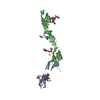 6eg0C 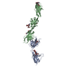 6eg1C 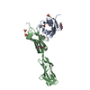 5eo9S S: Starting model for refinement C: citing same article ( |
|---|---|
| Similar structure data |


-Links
 PDBj
PDBj


- Assembly
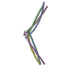
| Deposited unit | 
| ||||||||||
|---|---|---|---|---|---|---|---|---|---|---|---|
| 1 | 
| ||||||||||
| Unit cell |
|
-Components
| #1: Protein | Mass: 34503.098 Da / Num. of mol.: 1 / Fragment: UNP residues 40-341 Source method: isolated from a genetically manipulated source Source: (gene. exp.) Gene: DIP-alpha, 32791, CG13019, CG13020, Dmel\CG32791, CG32791, Dmel_CG32791 Production host:  Homo sapiens (human) / References: UniProt: Q9W4R3 Homo sapiens (human) / References: UniProt: Q9W4R3 |
|---|---|
| #2: Polysaccharide | 2-acetamido-2-deoxy-beta-D-glucopyranose-(1-4)-[alpha-L-fucopyranose-(1-6)]2-acetamido-2-deoxy-beta- ...2-acetamido-2-deoxy-beta-D-glucopyranose-(1-4)-[alpha-L-fucopyranose-(1-6)]2-acetamido-2-deoxy-beta-D-glucopyranose Source method: isolated from a genetically manipulated source |
| #3: Chemical | ChemComp-GOL /   Mass: 92.094 Da / Num. of mol.: 1 Mass: 92.094 Da / Num. of mol.: 1Source method: isolated from a genetically manipulated source Formula: C3H8O3 |
| #4: Water | ChemComp-HOH /  Mass: 18.015 Da / Num. of mol.: 5 / Source method: isolated from a natural source / Formula: H2O Mass: 18.015 Da / Num. of mol.: 5 / Source method: isolated from a natural source / Formula: H2O |
| Has protein modification | Y |
-Experimental details
-Experiment
| Experiment | Method: X-RAY DIFFRACTION / Number of used crystals: 1 |
|---|
- Sample preparation
| Crystal | Density Matthews: 4.77 Å3/Da / Density % sol: 74.22 % |
|---|---|
| Crystal grow | Temperature: 295 K / Method: vapor diffusion, hanging drop Details: 2% PEG3350, 17% Tacsimate, pH 7.0, 0.1 M HEPES, pH 7.0, cryoprotectant: 30% glycerol |
-Data collection
| Diffraction | Mean temperature: 100 K |
|---|---|
| Diffraction source | Source: SYNCHROTRON / Site: APS / Beamline: 24-ID-E / Wavelength: 0.979 Å |
| Detector | Type: ADSC QUANTUM 315 / Detector: CCD / Date: Apr 17, 2016 |
| Radiation | Monochromator: Cryogenically-cooled single crystal Si(220) side bounce Protocol: SINGLE WAVELENGTH / Monochromatic (M) / Laue (L): M / Scattering type: x-ray |
| Radiation wavelength | Wavelength: 0.979 Å / Relative weight: 1 |
| Reflection | Resolution: 2.9→67.78 Å / Num. obs: 14611 / % possible obs: 99.7 % / Redundancy: 4.6 % / Biso Wilson estimate: 79.9577746632 Å2 / Rmerge(I) obs: 0.081 / Net I/σ(I): 12.3 |
| Reflection shell | Resolution: 2.9→3.08 Å / Redundancy: 4.6 % / Rmerge(I) obs: 0.784 / Num. unique obs: 2349 / % possible all: 99.9 |
- Processing
| Software |
| ||||||||||||||||||||||||||||||||||||||||||
|---|---|---|---|---|---|---|---|---|---|---|---|---|---|---|---|---|---|---|---|---|---|---|---|---|---|---|---|---|---|---|---|---|---|---|---|---|---|---|---|---|---|---|---|
| Refinement | Method to determine structure: MOLECULAR REPLACEMENT Starting model: PDB entry 5EO9 Resolution: 2.9→19.938 Å / SU ML: 0.49 / Cross valid method: FREE R-VALUE / σ(F): 1.34 / Phase error: 32.57
| ||||||||||||||||||||||||||||||||||||||||||
| Solvent computation | Shrinkage radii: 0.9 Å / VDW probe radii: 1.11 Å | ||||||||||||||||||||||||||||||||||||||||||
| Refinement step | Cycle: LAST / Resolution: 2.9→19.938 Å
| ||||||||||||||||||||||||||||||||||||||||||
| Refine LS restraints |
| ||||||||||||||||||||||||||||||||||||||||||
| LS refinement shell |
|
