 Movie
Movie Controller

[English] 日本語
 Yorodumi
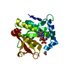
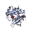
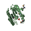
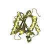
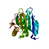
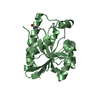
- PDB-6edv: Structure of a GNAT superfamily acetyltransferase PA3944 in compl... -
+ Open data

- Basic information
| Entry | Database: PDB / ID: 6edv | ||||||
|---|---|---|---|---|---|---|---|
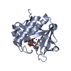
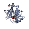
| Title | Structure of a GNAT superfamily acetyltransferase PA3944 in complex with CoA | ||||||
 Components Components | Acetyltransferase PA3944 | ||||||
 Keywords Keywords | TRANSFERASE / Gcn5-related N-acetyltransferase / GNAT / Pseudomonas aeruginosa / Structural Genomics / PSI-Biology / Center for Structural Genomics of Infectious Diseases / CSGID | ||||||
| Function / homology | : / Acetyltransferase (GNAT) domain / acyltransferase activity, transferring groups other than amino-acyl groups / Gcn5-related N-acetyltransferase (GNAT) domain profile. / GNAT domain / Acyl-CoA N-acyltransferase / Transferases; Acyltransferases; Transferring groups other than aminoacyl groups / COENZYME A / Acetyltransferase PA3944 Function and homology information Function and homology information | ||||||
| Biological species |   Pseudomonas aeruginosa (bacteria) Pseudomonas aeruginosa (bacteria) | ||||||
| Method |  X-RAY DIFFRACTION / SYNCHROTRON / MOLECULAR REPLACEMENT / Resolution: 1.35 Å X-RAY DIFFRACTION / SYNCHROTRON / MOLECULAR REPLACEMENT / Resolution: 1.35 Å | ||||||
 Authors Authors | Majorek, K.A. / Satchell, K.J.F. / Joachimiak, A. / Minor, W. / Center for Structural Genomics of Infectious Diseases (CSGID) | ||||||
 Citation Citation | Journal: Biochemistry / Year: 2018 Title: A Gcn5-Related N-Acetyltransferase (GNAT) Capable of Acetylating Polymyxin B and Colistin Antibiotics in Vitro. Authors: Czub, M.P. / Zhang, B. / Chiarelli, M.P. / Majorek, K.A. / Joe, L. / Porebski, P.J. / Revilla, A. / Wu, W. / Becker, D.P. / Minor, W. / Kuhn, M.L. | ||||||
| History |
|
- Structure visualization
| Structure viewer | Molecule: MolmilJmol/JSmol |
|---|
- Downloads & links
-Download
| PDBx/mmCIF format | 6edv.cif.gz | 99.7 KB | Display | PDBx/mmCIF format |
|---|---|---|---|---|
| PDB format | pdb6edv.ent.gz | 73.4 KB | Display | PDB format |
| PDBx/mmJSON format | 6edv.json.gz | Tree view | PDBx/mmJSON format | |
| Others |  Other downloads Other downloads |
-Validation report
| Arichive directory | https://data.pdbj.org/pub/pdb/validation_reports/ed/6edvftp://data.pdbj.org/pub/pdb/validation_reports/ed/6edv | HTTPS FTP |
|---|
-Related structure data
| Related structure data | 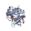 6eddC 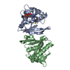 3fbuS C: citing same article ( S: Starting model for refinement |
|---|---|
| Similar structure data | |
| Other databases |


-Links
 PDBj
PDBj


- Assembly
| Deposited unit | 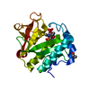
| ||||||||
|---|---|---|---|---|---|---|---|---|---|
| 1 |
| ||||||||
| Unit cell |
|
-Components
| #1: Protein | Mass: 22080.045 Da / Num. of mol.: 1 Source method: isolated from a genetically manipulated source Source: (gene. exp.) Pseudomonas aeruginosa (strain ATCC 15692 / DSM 22644 / CIP 104116 / JCM 14847 / LMG 12228 / 1C / PRS 101 / PAO1) (bacteria)Strain: ATCC 15692 / DSM 22644 / CIP 104116 / JCM 14847 / LMG 12228 / 1C / PRS 101 / PAO1 Gene: PA3944 / Production host: References: UniProt: Q9HX72, Transferases; Acyltransferases; Transferring groups other than aminoacyl groups | ||||
|---|---|---|---|---|---|
| #2: Chemical | ChemComp-CA /   Mass: 40.078 Da / Num. of mol.: 1 / Source method: obtained synthetically / Formula: Ca Mass: 40.078 Da / Num. of mol.: 1 / Source method: obtained synthetically / Formula: Ca | ||||
| #3: Chemical |   Mass: 62.068 Da / Num. of mol.: 2 / Source method: obtained synthetically / Formula: C2H6O2 Mass: 62.068 Da / Num. of mol.: 2 / Source method: obtained synthetically / Formula: C2H6O2#4: Chemical | ChemComp-COA / |   Mass: 767.534 Da / Num. of mol.: 1 / Source method: obtained synthetically / Formula: C21H36N7O16P3S Mass: 767.534 Da / Num. of mol.: 1 / Source method: obtained synthetically / Formula: C21H36N7O16P3S#5: Water | ChemComp-HOH / |  Mass: 18.015 Da / Num. of mol.: 155 / Source method: isolated from a natural source / Formula: H2O Mass: 18.015 Da / Num. of mol.: 155 / Source method: isolated from a natural source / Formula: H2O |
-Experimental details
-Experiment
| Experiment | Method: X-RAY DIFFRACTION / Number of used crystals: 1 |
|---|
- Sample preparation
| Crystal | Density Matthews: 2.02 Å3/Da / Density % sol: 39.09 % |
|---|---|
| Crystal grow | Temperature: 293 K / Method: vapor diffusion, sitting drop / pH: 7 Details: 100 mM Tris-HCl pH 7.0, 200 mM calcium acetate monohydrate, 20%w/v PEG 3000 |
-Data collection
| Diffraction | Mean temperature: 100 K |
|---|---|
| Diffraction source | Source: SYNCHROTRON / Site: APS  / Beamline: 19-ID / Wavelength: 0.979 Å / Beamline: 19-ID / Wavelength: 0.979 Å |
| Detector | Type: ADSC QUANTUM 315r / Detector: CCD / Date: Mar 3, 2012 / Details: mirrors |
| Radiation | Monochromator: Si 111 channel / Protocol: SINGLE WAVELENGTH / Monochromatic (M) / Laue (L): M / Scattering type: x-ray |
| Radiation wavelength | Wavelength: 0.979 Å / Relative weight: 1 |
| Reflection | Resolution: 1.35→30 Å / Num. obs: 40378 / % possible obs: 100 % / Redundancy: 13.8 % / Rmerge(I) obs: 0.122 / Net I/σ(I): 7.9 |
| Reflection shell | Resolution: 1.35→1.37 Å / Redundancy: 4.6 % / Rmerge(I) obs: 0.778 / % possible all: 99.7 |
- Processing
| Software |
| ||||||||||||||||||||
|---|---|---|---|---|---|---|---|---|---|---|---|---|---|---|---|---|---|---|---|---|---|
| Refinement | Method to determine structure: MOLECULAR REPLACEMENT Starting model: 3FBU Resolution: 1.35→30 Å / SU B: 1.457 / SU ML: 0.029 / Cross valid method: THROUGHOUT / σ(F): 0 / ESU R: 0.05 / ESU R Free: 0.047 / Details: HYDROGENS HAVE BEEN ADDED IN THE RIDING
| ||||||||||||||||||||
| Displacement parameters | Biso mean: 18.45 Å2
| ||||||||||||||||||||
| Refinement step | Cycle: LAST / Resolution: 1.35→30 Å
|
