 Movie
Movie Controller


+ Open data

- Basic information
| Entry | Database: PDB / ID: 6e3d | |||||||||
|---|---|---|---|---|---|---|---|---|---|---|
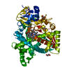
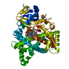
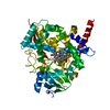
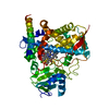
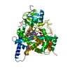
| Title | Atomic structure of Mycobacterium tuberculosis DppA | |||||||||
 Components Components |
| |||||||||
 Keywords Keywords | METAL BINDING PROTEIN / heme-binding / periplasmic protein | |||||||||
| Function / homology |  Function and homology information Function and homology informationpeptide transport / peptide transmembrane transporter activity / ATP-binding cassette (ABC) transporter complex / periplasmic space Similarity search - Function | |||||||||
| Biological species |   Mycobacterium tuberculosis (bacteria) Mycobacterium tuberculosis (bacteria) | |||||||||
| Method |  X-RAY DIFFRACTION / SYNCHROTRON / MOLECULAR REPLACEMENT / Resolution: 1.271 Å X-RAY DIFFRACTION / SYNCHROTRON / MOLECULAR REPLACEMENT / Resolution: 1.271 Å | |||||||||
 Authors Authors | Ko, Y. / Mitra, A. / Niederweis, M. / Cingolani, G. | |||||||||
| Funding support |  United States, 2items United States, 2items
| |||||||||
 Citation Citation | Journal: Nat Commun / Year: 2019 Title: Heme and hemoglobin utilization by Mycobacterium tuberculosis. Authors: Mitra, A. / Ko, Y.H. / Cingolani, G. / Niederweis, M. | |||||||||
| History |
|
- Structure visualization
| Structure viewer | Molecule: MolmilJmol/JSmol |
|---|
- Downloads & links
-Download
| PDBx/mmCIF format | 6e3d.cif.gz | 301.1 KB | Display | PDBx/mmCIF format |
|---|---|---|---|---|
| PDB format | pdb6e3d.ent.gz | 244.5 KB | Display | PDB format |
| PDBx/mmJSON format | 6e3d.json.gz | Tree view | PDBx/mmJSON format | |
| Others |  Other downloads Other downloads |
-Validation report
| Arichive directory | https://data.pdbj.org/pub/pdb/validation_reports/e3/6e3dftp://data.pdbj.org/pub/pdb/validation_reports/e3/6e3d | HTTPS FTP |
|---|
-Related structure data
| Related structure data |  6e4dC 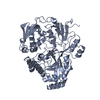 1b7hS S: Starting model for refinement C: citing same article ( |
|---|---|
| Similar structure data |


-Links
 PDBj
PDBj- Assembly
| Deposited unit | 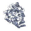
| ||||||||
|---|---|---|---|---|---|---|---|---|---|
| 1 |
| ||||||||
| Unit cell |
|
-Components
| #1: Protein | Mass: 55381.469 Da / Num. of mol.: 1 Source method: isolated from a genetically manipulated source Source: (gene. exp.) Mycobacterium tuberculosis (bacteria) / Gene: dppA, ERS007741_01322, ERS023446_02924 / Production host: |
|---|---|
| #2: Protein/peptide | Mass: 392.405 Da / Num. of mol.: 1 Source method: isolated from a genetically manipulated source Source: (gene. exp.) Mycobacterium tuberculosis (bacteria) / Production host: |
| #3: Water | ChemComp-HOH /  Mass: 18.015 Da / Num. of mol.: 566 / Source method: isolated from a natural source / Formula: H2O Mass: 18.015 Da / Num. of mol.: 566 / Source method: isolated from a natural source / Formula: H2O |
-Experimental details
-Experiment
| Experiment | Method: X-RAY DIFFRACTION / Number of used crystals: 1 |
|---|
- Sample preparation
| Crystal | Density Matthews: 1.95 Å3/Da / Density % sol: 36.84 % |
|---|---|
| Crystal grow | Temperature: 295 K / Method: vapor diffusion, hanging drop / pH: 6 / Details: 0.2 M Sodium Malonate pH 6.0 and 22% (w/v) PEG3350 |
-Data collection
| Diffraction | Mean temperature: 100 K |
|---|---|
| Diffraction source | Source: SYNCHROTRON / Site: SSRL / Beamline: BL9-2 / Wavelength: 0.97 Å |
| Detector | Type: DECTRIS PILATUS 6M / Detector: PIXEL / Date: Dec 21, 2017 |
| Radiation | Protocol: SINGLE WAVELENGTH / Monochromatic (M) / Laue (L): M / Scattering type: x-ray |
| Radiation wavelength | Wavelength: 0.97 Å / Relative weight: 1 |
| Reflection | Resolution: 1.27→50 Å / Num. obs: 102717 / % possible obs: 91.1 % / Redundancy: 6.1 % / Biso Wilson estimate: 13.5 Å2 / Rpim(I) all: 0.0028 / Rsym value: 0.0066 / Net I/σ(I): 37.8 |
| Reflection shell | Resolution: 1.27→1.32 Å / Redundancy: 3.2 % / Rpim(I) all: 0.029 / Rsym value: 0.0507 / % possible all: 51.3 |
- Processing
| Software |
| |||||||||||||||||||||||||||||||||||||||||||||||||||||||||||||||||||||||||||||||||||||||||||||||||||||||||
|---|---|---|---|---|---|---|---|---|---|---|---|---|---|---|---|---|---|---|---|---|---|---|---|---|---|---|---|---|---|---|---|---|---|---|---|---|---|---|---|---|---|---|---|---|---|---|---|---|---|---|---|---|---|---|---|---|---|---|---|---|---|---|---|---|---|---|---|---|---|---|---|---|---|---|---|---|---|---|---|---|---|---|---|---|---|---|---|---|---|---|---|---|---|---|---|---|---|---|---|---|---|---|---|---|---|---|
| Refinement | Method to determine structure: MOLECULAR REPLACEMENT Starting model: 1B7H Resolution: 1.271→38.007 Å / SU ML: 0.1 / Cross valid method: THROUGHOUT / σ(F): 0 / Phase error: 18.93
| |||||||||||||||||||||||||||||||||||||||||||||||||||||||||||||||||||||||||||||||||||||||||||||||||||||||||
| Solvent computation | Shrinkage radii: 0.9 Å / VDW probe radii: 1.11 Å | |||||||||||||||||||||||||||||||||||||||||||||||||||||||||||||||||||||||||||||||||||||||||||||||||||||||||
| Displacement parameters | Biso max: 99.61 Å2 / Biso mean: 22.2998 Å2 / Biso min: 9.58 Å2 | |||||||||||||||||||||||||||||||||||||||||||||||||||||||||||||||||||||||||||||||||||||||||||||||||||||||||
| Refinement step | Cycle: final / Resolution: 1.271→38.007 Å
| |||||||||||||||||||||||||||||||||||||||||||||||||||||||||||||||||||||||||||||||||||||||||||||||||||||||||
| LS refinement shell | Refine-ID: X-RAY DIFFRACTION / Rfactor Rfree error: 0 / Total num. of bins used: 14
|
