 Movie
Movie Controller


+ Open data

- Basic information
| Entry | Database: PDB / ID: 6e1n | |||||||||
|---|---|---|---|---|---|---|---|---|---|---|
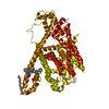
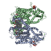
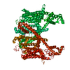
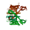
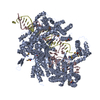
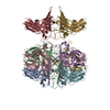
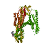
| Title | Structure of AtTPC1(DDE) in state 1 | |||||||||
 Components Components | Two pore calcium channel protein 1 | |||||||||
 Keywords Keywords | MEMBRANE PROTEIN / Two-pore channel | |||||||||
| Function / homology |  Function and homology information Function and homology informationregulation of jasmonic acid biosynthetic process / seed germination / regulation of stomatal movement / plant-type vacuole / vacuole / vacuolar membrane / monoatomic ion channel complex / voltage-gated calcium channel activity / calcium-mediated signaling / calcium channel activity ...regulation of jasmonic acid biosynthetic process / seed germination / regulation of stomatal movement / plant-type vacuole / vacuole / vacuolar membrane / monoatomic ion channel complex / voltage-gated calcium channel activity / calcium-mediated signaling / calcium channel activity / calcium ion transport / calcium ion binding / Golgi apparatus / identical protein binding / plasma membrane / cytosol Similarity search - Function | |||||||||
| Biological species |  | |||||||||
| Method | ELECTRON MICROSCOPY / single particle reconstruction / cryo EM / Resolution: 3.7 Å | |||||||||
 Authors Authors | Kintzer, A.F. / Green, E.M. / Cheng, Y. / Stroud, R.M. | |||||||||
| Funding support |  United States, 2items United States, 2items
| |||||||||
 Citation Citation | Journal: Proc Natl Acad Sci U S A / Year: 2018 Title: Structural basis for activation of voltage sensor domains in an ion channel TPC1. Authors: Alexander F Kintzer / Evan M Green / Pawel K Dominik / Michael Bridges / Jean-Paul Armache / Dawid Deneka / Sangwoo S Kim / Wayne Hubbell / Anthony A Kossiakoff / Yifan Cheng / Robert M Stroud / Abstract: Voltage-sensing domains (VSDs) couple changes in transmembrane electrical potential to conformational changes that regulate ion conductance through a central channel. Positively charged amino acids ...Voltage-sensing domains (VSDs) couple changes in transmembrane electrical potential to conformational changes that regulate ion conductance through a central channel. Positively charged amino acids inside each sensor cooperatively respond to changes in voltage. Our previous structure of a TPC1 channel captured an example of a resting-state VSD in an intact ion channel. To generate an activated-state VSD in the same channel we removed the luminal inhibitory Ca-binding site (Ca), which shifts voltage-dependent opening to more negative voltage and activation at 0 mV. Cryo-EM reveals two coexisting structures of the VSD, an intermediate state 1 that partially closes access to the cytoplasmic side but remains occluded on the luminal side and an intermediate activated state 2 in which the cytoplasmic solvent access to the gating charges closes, while luminal access partially opens. Activation can be thought of as moving a hydrophobic insulating region of the VSD from the external side to an alternate grouping on the internal side. This effectively moves the gating charges from the inside potential to that of the outside. Activation also requires binding of Ca to a cytoplasmic site (Ca). An X-ray structure with Ca removed and a near-atomic resolution cryo-EM structure with Ca removed define how dramatic conformational changes in the cytoplasmic domains may communicate with the VSD during activation. Together four structures provide a basis for understanding the voltage-dependent transition from resting to activated state, the tuning of VSD by thermodynamic stability, and this channel's requirement of cytoplasmic Ca ions for activation. | |||||||||
| History |
|
- Structure visualization
| Movie |
Movie viewer |
|---|---|
| Structure viewer | Molecule: MolmilJmol/JSmol |
- Downloads & links
-Download
| PDBx/mmCIF format | 6e1n.cif.gz | 249 KB | Display | PDBx/mmCIF format |
|---|---|---|---|---|
| PDB format | pdb6e1n.ent.gz | 198.4 KB | Display | PDB format |
| PDBx/mmJSON format | 6e1n.json.gz | Tree view | PDBx/mmJSON format | |
| Others |  Other downloads Other downloads |
-Validation report
| Arichive directory | https://data.pdbj.org/pub/pdb/validation_reports/e1/6e1nftp://data.pdbj.org/pub/pdb/validation_reports/e1/6e1n | HTTPS FTP |
|---|
-Related structure data
| Related structure data |  8958MC  8956C  8957C  8960C  6cx0C  6e1kC  6e1mC  6e1pC C: citing same article ( M: map data used to model this data |
|---|---|
| Similar structure data |

-Links
 PDBj
PDBj


- Assembly
| Deposited unit | 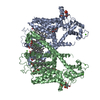
|
|---|---|
| 1 |
|
-Components
| #1: Protein | Mass: 84547.203 Da / Num. of mol.: 2 / Mutation: N240D, N454D, Q528E Source method: isolated from a genetically manipulated source Source: (gene. exp.)  #2: Chemical | ChemComp-CA /   Mass: 40.078 Da / Num. of mol.: 6 / Source method: obtained synthetically / Formula: Ca Mass: 40.078 Da / Num. of mol.: 6 / Source method: obtained synthetically / Formula: Ca#3: Chemical | ChemComp-PLM /   Mass: 256.424 Da / Num. of mol.: 16 / Source method: obtained synthetically / Formula: C16H32O2 Mass: 256.424 Da / Num. of mol.: 16 / Source method: obtained synthetically / Formula: C16H32O2#4: Chemical |   Mass: 704.998 Da / Num. of mol.: 2 / Source method: obtained synthetically / Formula: C39H77O8P Mass: 704.998 Da / Num. of mol.: 2 / Source method: obtained synthetically / Formula: C39H77O8P#5: Water | ChemComp-HOH / |  Mass: 18.015 Da / Num. of mol.: 2 / Source method: isolated from a natural source / Formula: H2O Mass: 18.015 Da / Num. of mol.: 2 / Source method: isolated from a natural source / Formula: H2OHas protein modification | Y | |
|---|
-Experimental details
-Experiment
| Experiment | Method: ELECTRON MICROSCOPY |
|---|---|
| EM experiment | Aggregation state: PARTICLE / 3D reconstruction method: single particle reconstruction |
- Sample preparation
| Component | Name: AtTPC1(DDE) / Type: COMPLEX / Entity ID: #1 / Source: MULTIPLE SOURCES |
|---|---|
| Source (natural) | Organism: |
| Buffer solution | pH: 7.3 |
| Specimen | Conc.: 0.8 mg/ml / Embedding applied: NO / Shadowing applied: NO / Staining applied: NO / Vitrification applied: YES / Details: Sample reconstituted into saposin A. |
| Vitrification | Instrument: FEI VITROBOT MARK III / Cryogen name: ETHANE / Humidity: 100 % / Chamber temperature: 20 K |
- Electron microscopy imaging
| Experimental equipment |  Model: Tecnai Polara / Image courtesy: FEI Company |
|---|---|
| Microscopy | Model: FEI POLARA 300 |
| Electron gun | Electron source:  FIELD EMISSION GUN / Accelerating voltage: 300 kV / Illumination mode: FLOOD BEAM FIELD EMISSION GUN / Accelerating voltage: 300 kV / Illumination mode: FLOOD BEAM |
| Electron lens | Mode: BRIGHT FIELD / Nominal magnification: 31000 X / Calibrated magnification: 41132 X / Nominal defocus max: 2000 nm / Nominal defocus min: 800 nm / Cs: 2 mm / C2 aperture diameter: 70 µm / Alignment procedure: COMA FREE |
| Specimen holder | Cryogen: NITROGEN |
| Image recording | Average exposure time: 0.2 sec. / Electron dose: 1 e/Å2 / Detector mode: SUPER-RESOLUTION / Film or detector model: GATAN K2 SUMMIT (4k x 4k) / Num. of grids imaged: 2 / Num. of real images: 3408 |
| Image scans | Movie frames/image: 60 / Used frames/image: 2-60 |
- Processing
| EM software |
| ||||||||||||||||||||||||||||||||
|---|---|---|---|---|---|---|---|---|---|---|---|---|---|---|---|---|---|---|---|---|---|---|---|---|---|---|---|---|---|---|---|---|---|
| CTF correction | Type: PHASE FLIPPING AND AMPLITUDE CORRECTION | ||||||||||||||||||||||||||||||||
| Particle selection | Num. of particles selected: 996035 | ||||||||||||||||||||||||||||||||
| Symmetry | Point symmetry: C2 (2 fold cyclic) | ||||||||||||||||||||||||||||||||
| 3D reconstruction | Resolution: 3.7 Å / Resolution method: FSC 0.143 CUT-OFF / Num. of particles: 44353 / Num. of class averages: 1 / Symmetry type: POINT | ||||||||||||||||||||||||||||||||
| Atomic model building | Protocol: RIGID BODY FIT / Space: REAL |
