 Movie
Movie Controller

[English] 日本語
 Yorodumi
- PDB-6e18: Crystal structure of Chlamydomonas reinhardtii HAP2 ectodomain pr... -
+ Open data

- Basic information
| Entry | Database: PDB / ID: 6.0E+18 | |||||||||
|---|---|---|---|---|---|---|---|---|---|---|
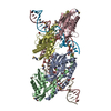
| Title | Crystal structure of Chlamydomonas reinhardtii HAP2 ectodomain provides structural insights of functional loops in green algae. | |||||||||
 Components Components | Hapless 2 | |||||||||
 Keywords Keywords | MEMBRANE PROTEIN / Gamete fusion / membrane binding motif / glycoprotein / cystine ladder | |||||||||
| Function / homology |  Function and homology information Function and homology informationfusion of sperm to egg plasma membrane involved in single fertilization / cell projection membrane / protein insertion into membrane / cytoplasmic vesicle membrane / cytoplasmic vesicle / lipid binding / plasma membrane Similarity search - Function | |||||||||
| Biological species |   Chlamydomonas reinhardtii (plant) Chlamydomonas reinhardtii (plant) | |||||||||
| Method |  X-RAY DIFFRACTION / SYNCHROTRON / MOLECULAR REPLACEMENT / molecular replacement / Resolution: 2.6 Å X-RAY DIFFRACTION / SYNCHROTRON / MOLECULAR REPLACEMENT / molecular replacement / Resolution: 2.6 Å | |||||||||
 Authors Authors | Baquero, E. / Legrand, P. / Rey, F.A. | |||||||||
| Funding support | European Union, 1items
| |||||||||
 Citation Citation | Journal: Structure / Year: 2019 Title: Species-Specific Functional Regions of the Green Alga Gamete Fusion Protein HAP2 Revealed by Structural Studies. Authors: Baquero, E. / Fedry, J. / Legrand, P. / Krey, T. / Rey, F.A. | |||||||||
| History |
|
- Structure visualization
| Structure viewer | Molecule: MolmilJmol/JSmol |
|---|
- Downloads & links
-Download
| PDBx/mmCIF format | 6e18.cif.gz | 225.2 KB | Display | PDBx/mmCIF format |
|---|---|---|---|---|
| PDB format | pdb6e18.ent.gz | 177.6 KB | Display | PDB format |
| PDBx/mmJSON format | 6e18.json.gz | Tree view | PDBx/mmJSON format | |
| Others |  Other downloads Other downloads |
-Validation report
| Arichive directory | https://data.pdbj.org/pub/pdb/validation_reports/e1/6e18ftp://data.pdbj.org/pub/pdb/validation_reports/e1/6e18 | HTTPS FTP |
|---|
-Related structure data
| Related structure data |  5mf1S S: Starting model for refinement |
|---|---|
| Similar structure data |

-Links
 PDBj
PDBj- Assembly
| Deposited unit | 
| ||||||||
|---|---|---|---|---|---|---|---|---|---|
| 1 | 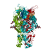
| ||||||||
| 2 | x 6
| ||||||||
| Unit cell |
| ||||||||
| Components on special symmetry positions |
|
-Components
-Protein , 1 types, 1 molecules A
| #1: Protein | Mass: 61432.645 Da / Num. of mol.: 1 Source method: isolated from a genetically manipulated source Source: (gene. exp.) Chlamydomonas reinhardtii (plant) / Gene: HAP2, GCS1 / Cell line (production host): Schneider S2 cells / Production host:  |
|---|
-Sugars , 3 types, 4 molecules 
| #2: Polysaccharide | 2-acetamido-2-deoxy-beta-D-glucopyranose-(1-4)-2-acetamido-2-deoxy-beta-D-glucopyranose Source method: isolated from a genetically manipulated source |
|---|---|
| #3: Polysaccharide | alpha-D-mannopyranose-(1-4)-2-acetamido-2-deoxy-beta-D-glucopyranose-(1-4)-[alpha-L-fucopyranose-(1- ...alpha-D-mannopyranose-(1-4)-2-acetamido-2-deoxy-beta-D-glucopyranose-(1-4)-[alpha-L-fucopyranose-(1-6)]2-acetamido-2-deoxy-beta-D-glucopyranose Source method: isolated from a genetically manipulated source |
| #4: Sugar |  Type: D-saccharide, beta linking / Mass: 221.208 Da / Num. of mol.: 2 Type: D-saccharide, beta linking / Mass: 221.208 Da / Num. of mol.: 2Source method: isolated from a genetically manipulated source Formula: C8H15NO6 |
-Non-polymers , 2 types, 84 molecules 

| #5: Chemical | ChemComp-GOL /  Mass: 92.094 Da / Num. of mol.: 1 / Source method: obtained synthetically / Formula: C3H8O3 Mass: 92.094 Da / Num. of mol.: 1 / Source method: obtained synthetically / Formula: C3H8O3 |
|---|---|
| #6: Water | ChemComp-HOH / Mass: 18.015 Da / Num. of mol.: 83 / Source method: isolated from a natural source / Formula: H2O |
-Details
| Has protein modification | Y |
|---|
-Experimental details
-Experiment
| Experiment | Method: X-RAY DIFFRACTION / Number of used crystals: 1 |
|---|
- Sample preparation
| Crystal | Density Matthews: 6.91 Å3/Da / Density % sol: 80 % / Description: Small hexagonal plates Preparation: The sample used for crystallization was a complex of Chlamydomonas reinhardtii HAP2 ectodomain with an antibody fragment scFv. However we could not reveal the presence of the scFv in our ...Preparation: The sample used for crystallization was a complex of Chlamydomonas reinhardtii HAP2 ectodomain with an antibody fragment scFv. However we could not reveal the presence of the scFv in our electron density maps. There are two possible explanations: 1) The bound scFv was displaced due to crystallization conditions/packing or 2) The scFv is still bound but disordered because is interacting with one of the disordered loops located in the large solvent volumes between the protomers. In this last case, it is expected that the solvent content of the crystal would be lower than the one currently reported |
|---|---|
| Crystal grow | Temperature: 291 K / Method: vapor diffusion, sitting drop / pH: 8.5 Details: 0.2M LiSO4, 0.1M Tris-HCl pH 8.5 and 25% w/v PEG 5000 MME |
-Data collection
| Diffraction | Mean temperature: 100 K | ||||||||||||||||||||||||
|---|---|---|---|---|---|---|---|---|---|---|---|---|---|---|---|---|---|---|---|---|---|---|---|---|---|
| Diffraction source | Source: SYNCHROTRON / Site: SOLEIL  / Beamline: PROXIMA 1 / Wavelength: 0.9786 Å / Beamline: PROXIMA 1 / Wavelength: 0.9786 Å | ||||||||||||||||||||||||
| Detector | Type: DECTRIS PILATUS3 S 6M / Detector: PIXEL / Date: Feb 4, 2018 | ||||||||||||||||||||||||
| Radiation | Protocol: SINGLE WAVELENGTH / Monochromatic (M) / Laue (L): M / Scattering type: x-ray | ||||||||||||||||||||||||
| Radiation wavelength | Wavelength: 0.9786 Å / Relative weight: 1 | ||||||||||||||||||||||||
| Reflection | Resolution: 2.6→40.13 Å / Num. obs: 53971 / % possible obs: 99.9 % / Redundancy: 19.6 % / Biso Wilson estimate: 67.34 Å2 / CC1/2: 0.997 / Rmerge(I) obs: 0.0592 / Rpim(I) all: 0.091 / Rrim(I) all: 0.406 / Net I/σ(I): 8.2 | ||||||||||||||||||||||||
| Reflection shell | Diffraction-ID: 1
|
-Phasing
| Phasing | Method: molecular replacement |
|---|
- Processing
| Software |
| ||||||||||||||||||||||||||||||||||||||||||||||||||||||||||||||||||||||||||||||||||||||||||||||||||||||||||||
|---|---|---|---|---|---|---|---|---|---|---|---|---|---|---|---|---|---|---|---|---|---|---|---|---|---|---|---|---|---|---|---|---|---|---|---|---|---|---|---|---|---|---|---|---|---|---|---|---|---|---|---|---|---|---|---|---|---|---|---|---|---|---|---|---|---|---|---|---|---|---|---|---|---|---|---|---|---|---|---|---|---|---|---|---|---|---|---|---|---|---|---|---|---|---|---|---|---|---|---|---|---|---|---|---|---|---|---|---|---|
| Refinement | Method to determine structure: MOLECULAR REPLACEMENT Starting model: 5MF1 Resolution: 2.6→38.99 Å / Cor.coef. Fo:Fc: 0.903 / Cor.coef. Fo:Fc free: 0.891 / SU R Cruickshank DPI: 0.277 / Cross valid method: THROUGHOUT / σ(F): 0 / SU R Blow DPI: 0.281 / SU Rfree Blow DPI: 0.227 / SU Rfree Cruickshank DPI: 0.227 Details: Due to marked diffraction anisotropy we post-processed the scaled intensities with the STARANISO program that performs an ellipsoidal resolution cut-off. The classical completeness is 70% up ...Details: Due to marked diffraction anisotropy we post-processed the scaled intensities with the STARANISO program that performs an ellipsoidal resolution cut-off. The classical completeness is 70% up to 2.6A resolution, but the "ellipsoidal completeness" is 94.5% up to the same resolution (79% in the last ellipsoidal shell).
| ||||||||||||||||||||||||||||||||||||||||||||||||||||||||||||||||||||||||||||||||||||||||||||||||||||||||||||
| Displacement parameters | Biso max: 136.28 Å2 / Biso mean: 50.89 Å2 / Biso min: 21.23 Å2
| ||||||||||||||||||||||||||||||||||||||||||||||||||||||||||||||||||||||||||||||||||||||||||||||||||||||||||||
| Refine analyze | Luzzati coordinate error obs: 0.41 Å | ||||||||||||||||||||||||||||||||||||||||||||||||||||||||||||||||||||||||||||||||||||||||||||||||||||||||||||
| Refinement step | Cycle: final / Resolution: 2.6→38.99 Å
| ||||||||||||||||||||||||||||||||||||||||||||||||||||||||||||||||||||||||||||||||||||||||||||||||||||||||||||
| Refine LS restraints |
| ||||||||||||||||||||||||||||||||||||||||||||||||||||||||||||||||||||||||||||||||||||||||||||||||||||||||||||
| LS refinement shell | Resolution: 2.6→2.67 Å / Rfactor Rfree error: 0 / Total num. of bins used: 19
| ||||||||||||||||||||||||||||||||||||||||||||||||||||||||||||||||||||||||||||||||||||||||||||||||||||||||||||
| Refinement TLS params. | Method: refined / Origin x: -51.3026 Å / Origin y: 26.2073 Å / Origin z: -25.4354 Å
| ||||||||||||||||||||||||||||||||||||||||||||||||||||||||||||||||||||||||||||||||||||||||||||||||||||||||||||
| Refinement TLS group |
|
