 Movie
Movie Controller

[English] 日本語
 Yorodumi
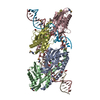
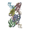
- PDB-7k2t: Mg2+/ATP-bound structure of the full-length WzmWzt O antigen ABC ... -
+ Open data

- Basic information
| Entry | Database: PDB / ID: 7k2t | ||||||
|---|---|---|---|---|---|---|---|
| Title | Mg2+/ATP-bound structure of the full-length WzmWzt O antigen ABC transporter in lipid nanodiscs | ||||||
 Components Components |
| ||||||
 Keywords Keywords | TRANSPORT PROTEIN / O antigen transporter / integral membrane protein / lipopolysaccharide LPS biosynthesis | ||||||
| Function / homology |  Function and homology information Function and homology informationpolysaccharide transport / lipopolysaccharide transport / ABC-type transporter activity / ATP hydrolysis activity / ATP binding / membrane / plasma membrane Similarity search - Function | ||||||
| Biological species |   Aquifex aeolicus (bacteria) Aquifex aeolicus (bacteria) | ||||||
| Method | ELECTRON MICROSCOPY / single particle reconstruction / cryo EM / Resolution: 3.6 Å | ||||||
 Authors Authors | Caffalette, C.A. / Zimmer, J. | ||||||
| Funding support |  United States, 1items United States, 1items
| ||||||
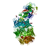
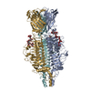
 Citation Citation | Journal: Proc Natl Acad Sci U S A / Year: 2021 Title: Cryo-EM structure of the full-length WzmWzt ABC transporter required for lipid-linked O antigen transport. Authors: Christopher A Caffalette / Jochen Zimmer / Abstract: O antigens are important cell surface polysaccharides in gram-negative bacteria where they extend core lipopolysaccharides in the extracellular leaflet of the outer membrane. O antigen structures are ...O antigens are important cell surface polysaccharides in gram-negative bacteria where they extend core lipopolysaccharides in the extracellular leaflet of the outer membrane. O antigen structures are serotype specific and form extended cell surface barriers endowing many pathogens with survival benefits. In the ABC transporter-dependent biosynthesis pathway, O antigens are assembled on the cytosolic side of the inner membrane on a lipid anchor and reoriented to the periplasmic leaflet by the channel-forming WzmWzt ABC transporter for ligation to the core lipopolysaccharides. In many cases, this process depends on the chemical modification of the O antigen's nonreducing terminus, sensed by WzmWzt via a carbohydrate-binding domain (CBD) that extends its nucleotide-binding domain (NBD). Here, we provide the cryo-electron microscopy structure of the full-length WzmWzt transporter from bound to adenosine triphosphate (ATP) and in a lipid environment, revealing a highly asymmetric transporter organization. The CBDs dimerize and associate with only one NBD. Conserved loops at the CBD dimer interface straddle a conserved peripheral NBD helix. The CBD dimer is oriented perpendicularly to the NBDs and its putative ligand-binding sites face the transporter to likely modulate ATPase activity upon O antigen binding. Further, our structure reveals a closed WzmWzt conformation in which an aromatic belt near the periplasmic channel exit seals the transporter in a resting, ATP-bound state. The sealed transmembrane channel is asymmetric, with one open and one closed cytosolic and periplasmic portal. The structure provides important insights into O antigen recruitment to and translocation by WzmWzt and related ABC transporters. | ||||||
| History |
|
- Structure visualization
| Movie |
Movie viewer |
|---|---|
| Structure viewer | Molecule: MolmilJmol/JSmol |
- Downloads & links
-Download
| PDBx/mmCIF format | 7k2t.cif.gz | 231 KB | Display | PDBx/mmCIF format |
|---|---|---|---|---|
| PDB format | pdb7k2t.ent.gz | 185.8 KB | Display | PDB format |
| PDBx/mmJSON format | 7k2t.json.gz | Tree view | PDBx/mmJSON format | |
| Others |  Other downloads Other downloads |
-Validation report
| Arichive directory | https://data.pdbj.org/pub/pdb/validation_reports/k2/7k2tftp://data.pdbj.org/pub/pdb/validation_reports/k2/7k2t | HTTPS FTP |
|---|
-Related structure data
| Related structure data |  22644MC M: map data used to model this data C: citing same article ( |
|---|---|
| Similar structure data | |
| EM raw data | EMPIAR-10558 (Title: Cryo EM strutcture of the full-length WzmWzt ABC transporter required for lipid-linked O antigen transport Data size: 2.2 TB Data #1: Unaligned multi-frame micrograph [micrographs - multiframe]) |


-Links
 PDBj
PDBj

- Assembly
| Deposited unit | 
|
|---|---|
| 1 |
|
-Components
| #1: Protein | Mass: 46283.426 Da / Num. of mol.: 2 / Mutation: E167Q Source method: isolated from a genetically manipulated source Source: (gene. exp.) Aquifex aeolicus (strain VF5) (bacteria)Strain: VF5 / Gene: abcT4, aq_1094 / Production host: #2: Protein | Mass: 30027.871 Da / Num. of mol.: 2 Source method: isolated from a genetically manipulated source Source: (gene. exp.) Aquifex aeolicus (strain VF5) (bacteria)Strain: VF5 / Gene: abcT3, aq_1095 / Production host: #3: Chemical |   Mass: 507.181 Da / Num. of mol.: 2 / Source method: obtained synthetically / Formula: C10H16N5O13P3 / Feature type: SUBJECT OF INVESTIGATION / Comment: ATP, energy-carrying molecule*YM Mass: 507.181 Da / Num. of mol.: 2 / Source method: obtained synthetically / Formula: C10H16N5O13P3 / Feature type: SUBJECT OF INVESTIGATION / Comment: ATP, energy-carrying molecule*YM#4: Chemical |   Mass: 24.305 Da / Num. of mol.: 2 / Source method: obtained synthetically / Formula: Mg / Feature type: SUBJECT OF INVESTIGATION Mass: 24.305 Da / Num. of mol.: 2 / Source method: obtained synthetically / Formula: Mg / Feature type: SUBJECT OF INVESTIGATIONHas ligand of interest | Y | |
|---|
-Experimental details
-Experiment
| Experiment | Method: ELECTRON MICROSCOPY |
|---|---|
| EM experiment | Aggregation state: PARTICLE / 3D reconstruction method: single particle reconstruction |
- Sample preparation
| Component | Name: Full-length WzmWzt O antigen ABC transporter / Type: COMPLEX / Entity ID: #1-#2 / Source: RECOMBINANT |
|---|---|
| Molecular weight | Value: 0.152385 MDa / Experimental value: NO |
| Source (natural) | Organism: Aquifex aeolicus VF5 (bacteria) |
| Source (recombinant) | Organism: |
| Buffer solution | pH: 7.5 |
| Specimen | Conc.: 1.1 mg/ml / Embedding applied: NO / Shadowing applied: NO / Staining applied: NO / Vitrification applied: YES / Details: 2.5 mM ATP and 2.5 mM MgCl2 |
| Specimen support | Grid material: COPPER / Grid mesh size: 300 divisions/in. / Grid type: Quantifoil R1.2/1.3 |
| Vitrification | Instrument: FEI VITROBOT MARK IV / Cryogen name: ETHANE / Humidity: 100 % / Chamber temperature: 277.15 K |
- Electron microscopy imaging
| Experimental equipment |  Model: Titan Krios / Image courtesy: FEI Company |
|---|---|
| Microscopy | Model: FEI TITAN KRIOS |
| Electron gun | Electron source:  FIELD EMISSION GUN / Accelerating voltage: 300 kV / Illumination mode: OTHER FIELD EMISSION GUN / Accelerating voltage: 300 kV / Illumination mode: OTHER |
| Electron lens | Mode: BRIGHT FIELD / Nominal magnification: 81000 X / Nominal defocus max: 2200 nm / Nominal defocus min: 1200 nm / Cs: 2.7 mm |
| Image recording | Average exposure time: 2.4767 sec. / Electron dose: 45.0252 e/Å2 / Film or detector model: GATAN K3 (6k x 4k) / Num. of grids imaged: 2 / Num. of real images: 5938 |
- Processing
| EM software |
| ||||||||||||||||||||||||||||||||||||||||||||
|---|---|---|---|---|---|---|---|---|---|---|---|---|---|---|---|---|---|---|---|---|---|---|---|---|---|---|---|---|---|---|---|---|---|---|---|---|---|---|---|---|---|---|---|---|---|
| CTF correction | Type: PHASE FLIPPING AND AMPLITUDE CORRECTION | ||||||||||||||||||||||||||||||||||||||||||||
| Symmetry | Point symmetry: C1 (asymmetric) | ||||||||||||||||||||||||||||||||||||||||||||
| 3D reconstruction | Resolution: 3.6 Å / Resolution method: FSC 0.143 CUT-OFF / Num. of particles: 48174 / Symmetry type: POINT | ||||||||||||||||||||||||||||||||||||||||||||
| Atomic model building | Protocol: AB INITIO MODEL / Space: REAL | ||||||||||||||||||||||||||||||||||||||||||||
| Atomic model building |
|


