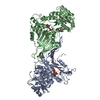
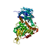
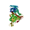
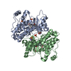
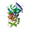
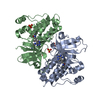
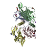
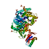
- PDB-6e08: Crystal structure of G6PD in complex with structural NADP -
+
Open data
ID or keywords:
Loading...
-
Basic information
Entry
Database: PDB / ID: 600000000
Title
Crystal structure of G6PD in complex with structural NADP
Components
Glucose-6-phosphate 1-dehydrogenase
Keywords
OXIDOREDUCTASE / G6PD / NADP
Function / homology
Function and homology information
pentose biosynthetic process / ribose phosphate biosynthetic process / response to iron(III) ion / positive regulation of calcium ion transmembrane transport via high voltage-gated calcium channel / glucose-6-phosphate dehydrogenase (NADP+) / glucose-6-phosphate dehydrogenase activity / Pentose phosphate pathway / pentose-phosphate shunt, oxidative branch / negative regulation of cell growth involved in cardiac muscle cell development / glucose 6-phosphate metabolic process ...pentose biosynthetic process / ribose phosphate biosynthetic process / response to iron(III) ion / positive regulation of calcium ion transmembrane transport via high voltage-gated calcium channel / glucose-6-phosphate dehydrogenase (NADP+) / glucose-6-phosphate dehydrogenase activity / Pentose phosphate pathway / pentose-phosphate shunt, oxidative branch / negative regulation of cell growth involved in cardiac muscle cell development / glucose 6-phosphate metabolic process / NADP+ metabolic process / pentose-phosphate shunt / D-glucose binding / NFE2L2 regulates pentose phosphate pathway genes / Oxidoreductases / cholesterol biosynthetic process / erythrocyte maturation / response to food / negative regulation of reactive oxygen species metabolic process / substantia nigra development / regulation of neuron apoptotic process / TP53 Regulates Metabolic Genes / lipid metabolic process / glutathione metabolic process / glucose metabolic process / cytoplasmic side of plasma membrane / centriolar satellite / NADP binding / cellular response to oxidative stress / response to ethanol / protein homodimerization activity / extracellular exosome / membrane / identical protein binding / cytoplasm / cytosol Similarity search - Function
Resolution: 1.9→18.28 Å / Cor.coef. Fo:Fc: 0.962 / Cor.coef. Fo:Fc free: 0.943 / SU B: 8.127 / SU ML: 0.104 / Cross valid method: THROUGHOUT / ESU R: 0.149 / ESU R Free: 0.139 / Details: HYDROGENS HAVE BEEN ADDED IN THE RIDING POSITIONS
Rfactor
Num. reflection
% reflection
Selection details
Rfree
0.21258
2171
5.1 %
RANDOM
Rwork
0.17182
-
-
-
obs
0.17402
40413
94.69 %
-
Solvent computation
Ion probe radii: 0.8 Å / Shrinkage radii: 0.8 Å / VDW probe radii: 1.2 Å
 Movie
Movie Controller


 Open data
 Basic information
 Components
 Keywords
 Function and homology information
 Homo sapiens (human)
 X-RAY DIFFRACTION /
  Authors
 Citation
 Structure visualization
 Downloads & links
 Other downloads
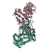


 PDBj
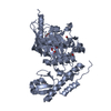
PDBj Assembly
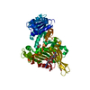


 Mass: 743.405 Da / Num. of mol.: 1 / Source method: obtained synthetically / Formula: C21H28N7O17P3
 Mass: 92.094 Da / Num. of mol.: 5 / Source method: obtained synthetically / Formula: C3H8O3
 Mass: 18.015 Da / Num. of mol.: 406 / Source method: isolated from a natural source / Formula: H2O
 Sample preparation
 / Beamline: BL12-2 / Wavelength: 1 Å
 Processing