 Movie
Movie Controller


+ Open data

- Basic information
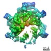
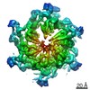
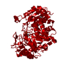
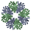
| Entry | Database: PDB / ID: 6ds5 | ||||||||||||||||||||||||||||||||||||||||||||||||||||||
|---|---|---|---|---|---|---|---|---|---|---|---|---|---|---|---|---|---|---|---|---|---|---|---|---|---|---|---|---|---|---|---|---|---|---|---|---|---|---|---|---|---|---|---|---|---|---|---|---|---|---|---|---|---|---|---|
| Title | Cryo EM structure of human SEIPIN | ||||||||||||||||||||||||||||||||||||||||||||||||||||||
 Components Components | Seipin | ||||||||||||||||||||||||||||||||||||||||||||||||||||||
 Keywords Keywords | LIPID BINDING PROTEIN / Lipid droplets / adipogenesis | ||||||||||||||||||||||||||||||||||||||||||||||||||||||
| Function / homology |  Function and homology information Function and homology informationlipid droplet formation / lipid storage / lipid droplet organization / negative regulation of lipid catabolic process / fat cell differentiation / lipid catabolic process / lipid droplet / phospholipid binding / positive regulation of cold-induced thermogenesis / endoplasmic reticulum membrane Similarity search - Function | ||||||||||||||||||||||||||||||||||||||||||||||||||||||
| Biological species |  Homo sapiens (human) Homo sapiens (human) | ||||||||||||||||||||||||||||||||||||||||||||||||||||||
| Method | ELECTRON MICROSCOPY / single particle reconstruction / cryo EM / Resolution: 3.8 Å | ||||||||||||||||||||||||||||||||||||||||||||||||||||||
 Authors Authors | Yan, R.H. / Qian, H.W. / Yan, N. / Yang, H.Y. | ||||||||||||||||||||||||||||||||||||||||||||||||||||||
| Funding support |  China, China,  Australia, 2items Australia, 2items
| ||||||||||||||||||||||||||||||||||||||||||||||||||||||
 Citation Citation | Journal: Dev Cell / Year: 2018 Title: Human SEIPIN Binds Anionic Phospholipids. Authors: Renhong Yan / Hongwu Qian / Ivan Lukmantara / Mingming Gao / Ximing Du / Nieng Yan / Hongyuan Yang /  Abstract: The biogenesis of lipid droplets (LDs) and the development of adipocytes are two key aspects of mammalian fat storage. SEIPIN, an integral membrane protein of the endoplasmic reticulum (ER), plays a ...The biogenesis of lipid droplets (LDs) and the development of adipocytes are two key aspects of mammalian fat storage. SEIPIN, an integral membrane protein of the endoplasmic reticulum (ER), plays a critical role in both LD formation and adipogenesis. The molecular function of SEIPIN, however, has yet to be elucidated. Here, we report the cryogenic electron microscopy structure of human SEIPIN at 3.8 Å resolution. SEIPIN exists as an undecamer, and this oligomerization state is critical for its physiological function. The evolutionarily conserved lumenal domain of SEIPIN forms an eight-stranded β sandwich fold. Both full-length SEIPIN and its lumenal domain can bind anionic phospholipids including phosphatidic acid. Our results suggest that SEIPIN forms a scaffold that helps maintain phospholipid homeostasis and surface tension of the ER. | ||||||||||||||||||||||||||||||||||||||||||||||||||||||
| History |
|
- Structure visualization
| Movie |
Movie viewer |
|---|---|
| Structure viewer | Molecule: MolmilJmol/JSmol |
- Downloads & links
-Download
| PDBx/mmCIF format | 6ds5.cif.gz | 347.1 KB | Display | PDBx/mmCIF format |
|---|---|---|---|---|
| PDB format | pdb6ds5.ent.gz | 266 KB | Display | PDB format |
| PDBx/mmJSON format | 6ds5.json.gz | Tree view | PDBx/mmJSON format | |
| Others |  Other downloads Other downloads |
-Validation report
| Arichive directory | https://data.pdbj.org/pub/pdb/validation_reports/ds/6ds5ftp://data.pdbj.org/pub/pdb/validation_reports/ds/6ds5 | HTTPS FTP |
|---|
-Related structure data
| Related structure data | 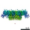 8909MC M: map data used to model this data C: citing same article ( |
|---|---|
| Similar structure data |

-Links
 PDBj
PDBj- Assembly
| Deposited unit | 
|
|---|---|
| 1 |
|
-Components
| #1: Protein | Mass: 45773.559 Da / Num. of mol.: 11 Source method: isolated from a genetically manipulated source Source: (gene. exp.) Homo sapiens (human) / Gene: BSCL2 / Production host: Homo sapiens (human) / References: UniProt: Q96G97#2: Polysaccharide | 2-acetamido-2-deoxy-beta-D-glucopyranose-(1-4)-2-acetamido-2-deoxy-beta-D-glucopyranose Source method: isolated from a genetically manipulated source Has protein modification | Y | |
|---|
-Experimental details
-Experiment
| Experiment | Method: ELECTRON MICROSCOPY |
|---|---|
| EM experiment | Aggregation state: PARTICLE / 3D reconstruction method: single particle reconstruction |
- Sample preparation
| Component | Name: SEIPIN / Type: ORGANELLE OR CELLULAR COMPONENT / Entity ID: #1 / Source: RECOMBINANT |
|---|---|
| Source (natural) | Organism: Homo sapiens (human) |
| Source (recombinant) | Organism: Homo sapiens (human) |
| Buffer solution | pH: 8 |
| Specimen | Conc.: 10 mg/ml / Embedding applied: NO / Shadowing applied: NO / Staining applied: NO / Vitrification applied: YES |
| Specimen support | Details: unspecified |
| Vitrification | Cryogen name: ETHANE |
- Electron microscopy imaging
| Experimental equipment |  Model: Titan Krios / Image courtesy: FEI Company |
|---|---|
| Microscopy | Model: FEI TITAN KRIOS |
| Electron gun | Electron source:  FIELD EMISSION GUN / Accelerating voltage: 300 kV / Illumination mode: FLOOD BEAM FIELD EMISSION GUN / Accelerating voltage: 300 kV / Illumination mode: FLOOD BEAM |
| Electron lens | Mode: BRIGHT FIELD |
| Image recording | Electron dose: 50 e/Å2 / Film or detector model: FEI FALCON II (4k x 4k) |
- Processing
| Software | Name: PHENIX / Version: 1.13_2998: / Classification: refinement | ||||||||||||||||||||||||
|---|---|---|---|---|---|---|---|---|---|---|---|---|---|---|---|---|---|---|---|---|---|---|---|---|---|
| EM software | Name: PHENIX / Category: model refinement | ||||||||||||||||||||||||
| CTF correction | Type: PHASE FLIPPING AND AMPLITUDE CORRECTION | ||||||||||||||||||||||||
| 3D reconstruction | Resolution: 3.8 Å / Resolution method: FSC 0.143 CUT-OFF / Num. of particles: 99281 / Symmetry type: POINT | ||||||||||||||||||||||||
| Refine LS restraints |
|
