 Movie
Movie Controller


+ Open data

- Basic information
| Entry | Database: EMDB / ID: EMD-8909 | |||||||||
|---|---|---|---|---|---|---|---|---|---|---|
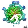
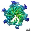
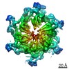
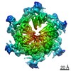
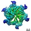
| Title | Cryo EM structure of human SEIPIN | |||||||||
 Map data Map data | Cryo EM structure of human SEIPIN, primary map | |||||||||
 Sample Sample |
| |||||||||
 Keywords Keywords | Lipid droplets / adipogenesis / LIPID BINDING PROTEIN | |||||||||
| Function / homology |  Function and homology information Function and homology informationlipid droplet formation / lipid storage / lipid droplet organization / negative regulation of lipid catabolic process / fat cell differentiation / lipid catabolic process / lipid droplet / phospholipid binding / positive regulation of cold-induced thermogenesis / endoplasmic reticulum membrane Similarity search - Function | |||||||||
| Biological species |  Homo sapiens (human) Homo sapiens (human) | |||||||||
| Method | single particle reconstruction / cryo EM / Resolution: 3.8 Å | |||||||||
 Authors Authors | Yan RH / Qian HW | |||||||||
| Funding support |  China, China,  Australia, 2 items Australia, 2 items
| |||||||||
 Citation Citation | Journal: Dev Cell / Year: 2018 Title: Human SEIPIN Binds Anionic Phospholipids. Authors: Renhong Yan / Hongwu Qian / Ivan Lukmantara / Mingming Gao / Ximing Du / Nieng Yan / Hongyuan Yang /  Abstract: The biogenesis of lipid droplets (LDs) and the development of adipocytes are two key aspects of mammalian fat storage. SEIPIN, an integral membrane protein of the endoplasmic reticulum (ER), plays a ...The biogenesis of lipid droplets (LDs) and the development of adipocytes are two key aspects of mammalian fat storage. SEIPIN, an integral membrane protein of the endoplasmic reticulum (ER), plays a critical role in both LD formation and adipogenesis. The molecular function of SEIPIN, however, has yet to be elucidated. Here, we report the cryogenic electron microscopy structure of human SEIPIN at 3.8 Å resolution. SEIPIN exists as an undecamer, and this oligomerization state is critical for its physiological function. The evolutionarily conserved lumenal domain of SEIPIN forms an eight-stranded β sandwich fold. Both full-length SEIPIN and its lumenal domain can bind anionic phospholipids including phosphatidic acid. Our results suggest that SEIPIN forms a scaffold that helps maintain phospholipid homeostasis and surface tension of the ER. | |||||||||
| History |
|
- Structure visualization
| Movie |
Movie viewer |
|---|---|
| Structure viewer | EM map: SurfViewMolmilJmol/JSmol |
| Supplemental images |

- Downloads & links
-EMDB archive
| Map data | emd_8909.map.gz | 164.6 MB | EMDB map data format | |
|---|---|---|---|---|
| Header (meta data) | emd-8909-v30.xmlemd-8909.xml | 16.2 KB 16.2 KB | Display Display | EMDB header |
| Images | 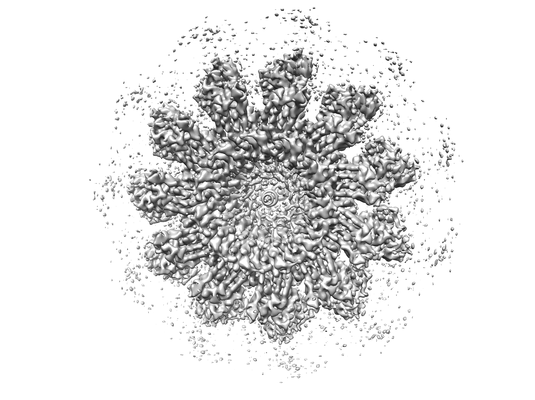 emd_8909.png emd_8909.png | 75.5 KB | ||
| Filedesc metadata | emd-8909.cif.gz | 6.6 KB | ||
| Archive directory |  http://ftp.pdbj.org/pub/emdb/structures/EMD-8909ftp://ftp.pdbj.org/pub/emdb/structures/EMD-8909 http://ftp.pdbj.org/pub/emdb/structures/EMD-8909ftp://ftp.pdbj.org/pub/emdb/structures/EMD-8909 | HTTPS FTP |
-Related structure data
| Related structure data | 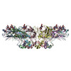 6ds5MC M: atomic model generated by this map C: citing same article ( |
|---|---|
| Similar structure data |

-Links
| EMDB pages | EMDB (EBI/PDBe) / EMDataResource |
|---|
-Map
| File | Download / File: emd_8909.map.gz / Format: CCP4 / Size: 178 MB / Type: IMAGE STORED AS FLOATING POINT NUMBER (4 BYTES) | ||||||||||||||||||||||||||||||||||||||||||||||||||||||||||||
|---|---|---|---|---|---|---|---|---|---|---|---|---|---|---|---|---|---|---|---|---|---|---|---|---|---|---|---|---|---|---|---|---|---|---|---|---|---|---|---|---|---|---|---|---|---|---|---|---|---|---|---|---|---|---|---|---|---|---|---|---|---|
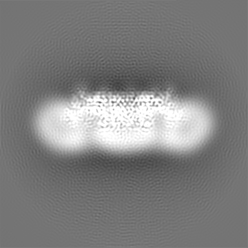
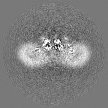
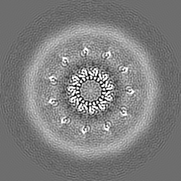
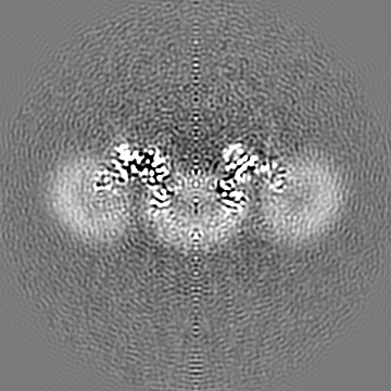
| Annotation | Cryo EM structure of human SEIPIN, primary map | ||||||||||||||||||||||||||||||||||||||||||||||||||||||||||||
| Projections & slices | Image control
Images are generated by Spider. | ||||||||||||||||||||||||||||||||||||||||||||||||||||||||||||
| Voxel size | X=Y=Z: 0.88 Å | ||||||||||||||||||||||||||||||||||||||||||||||||||||||||||||
| Density |
| ||||||||||||||||||||||||||||||||||||||||||||||||||||||||||||
| Symmetry | Space group: 1 | ||||||||||||||||||||||||||||||||||||||||||||||||||||||||||||
| Details | EMDB XML:
CCP4 map header:
| ||||||||||||||||||||||||||||||||||||||||||||||||||||||||||||
 Z (Sec.)
 Y (Row.)
 X (Col.)


-Supplemental data
- Sample components
-Entire : SEIPIN
| Entire | Name: SEIPIN |
|---|---|
| Components |
|
-Supramolecule #1: SEIPIN
| Supramolecule | Name: SEIPIN / type: organelle_or_cellular_component / ID: 1 / Parent: 0 / Macromolecule list: all |
|---|---|
| Source (natural) | Organism: Homo sapiens (human) |
-Macromolecule #1: Seipin
| Macromolecule | Name: Seipin / type: protein_or_peptide / ID: 1 / Number of copies: 11 / Enantiomer: LEVO |
|---|---|
| Source (natural) | Organism: Homo sapiens (human) |
| Molecular weight | Theoretical: 45.773559 KDa |
| Recombinant expression | Organism: Homo sapiens (human) |
| Sequence | String: MSGRWSHPQF EKVNDPPVPA LLWAQEVGQV LAGRARRLLL QFGVLFCTIL LLLWVSVFLY GSFYYSYMPT VSHLSPVHFY YRTDCDSST TSLCSFPVAN VSLTKGGRDR VLMYGQPYRV TLELELPESP VNQDLGMFLV TISCYTRGGR IISTSSRSVM L HYRSDLLQ ...String: MSGRWSHPQF EKVNDPPVPA LLWAQEVGQV LAGRARRLLL QFGVLFCTIL LLLWVSVFLY GSFYYSYMPT VSHLSPVHFY YRTDCDSST TSLCSFPVAN VSLTKGGRDR VLMYGQPYRV TLELELPESP VNQDLGMFLV TISCYTRGGR IISTSSRSVM L HYRSDLLQ MLDTLVFSSL LLFGFAEQKQ LLEVELYADY RENSYVPTTG AIIEIHSKRI QLYGAYLRIH AHFTGLRYLL YN FPMTCAF IGVASNFTFL SVIVLFSYMQ WVWGGIWPRH RFSLQVNIRK RDNSRKEVQR RISAHQPGPE GQEESTPQSD VTE DGESPE DPSGTEGQLS EEEKPDQQPL SGEEELEPEA SDGSGSWEDA ALLTEANLPA PAPASASAPV LETLGSSEPA GGAL RQRPT CSSS UniProtKB: Seipin |
-Experimental details
-Structure determination
| Method | cryo EM |
|---|---|
 Processing Processing | single particle reconstruction |
| Aggregation state | particle |
-Sample preparation
| Concentration | 10 mg/mL |
|---|---|
| Buffer | pH: 8 |
| Grid | Details: unspecified |
| Vitrification | Cryogen name: ETHANE |
- Electron microscopy
| Microscope | FEI TITAN KRIOS |
|---|---|
| Image recording | Film or detector model: FEI FALCON II (4k x 4k) / Average electron dose: 50.0 e/Å2 |
| Electron beam | Acceleration voltage: 300 kV / Electron source:  FIELD EMISSION GUN FIELD EMISSION GUN |
| Electron optics | Illumination mode: FLOOD BEAM / Imaging mode: BRIGHT FIELD |
| Experimental equipment |  Model: Titan Krios / Image courtesy: FEI Company |
