 Movie
Movie Controller

[English] 日本語
 Yorodumi
- PDB-6dcj: LpoA N-terminal domain from Haemophilus influenzae; monoclinic fo... -
+ Open data

- Basic information
| Entry | Database: PDB / ID: 6dcj | ||||||
|---|---|---|---|---|---|---|---|
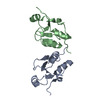
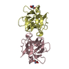
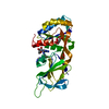
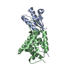
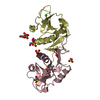
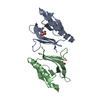
| Title | LpoA N-terminal domain from Haemophilus influenzae; monoclinic form at 1.35 A resolution | ||||||
 Components Components | Penicillin-binding protein activator LpoA | ||||||
 Keywords Keywords | BIOSYNTHETIC PROTEIN / peptidoglycan synthesis / TPR-like / outer membrane lipoprotein / PROTEIN BINDING | ||||||
| Function / homology |  Function and homology information Function and homology informationperiplasmic side of cell outer membrane / peptidoglycan biosynthetic process / enzyme regulator activity / regulation of cell shape Similarity search - Function | ||||||
| Biological species |  Haemophilus influenzae (bacteria) Haemophilus influenzae (bacteria) | ||||||
| Method |  X-RAY DIFFRACTION / SYNCHROTRON / MOLECULAR REPLACEMENT / molecular replacement / Resolution: 1.35 Å X-RAY DIFFRACTION / SYNCHROTRON / MOLECULAR REPLACEMENT / molecular replacement / Resolution: 1.35 Å | ||||||
 Authors Authors | Vijayalakshmi, J. / Saper, M.A. | ||||||
 Citation Citation | Journal: Acta Crystallogr.,Sect.F / Year: 2019 Title: Crystal structures of the amino-terminal domain of LpoA from Escherichia coli and Haemophilus influenzae. Authors: Kelley, A. / Vijayalakshmi, J. / Saper, M.A. #1: Journal: J. Biol. Chem. / Year: 2017Title: Structural analyses of the Haemophilus influenzae peptidoglycan synthase activator LpoA suggest multiple conformations in solution Authors: Sathiyamoorthy, K. / Vijayalakshmi, J. / Tirupati, B. / Fan, L. / Saper, M.A. | ||||||
| History |
|
- Structure visualization
| Structure viewer | Molecule: MolmilJmol/JSmol |
|---|
- Downloads & links
-Download
| PDBx/mmCIF format | 6dcj.cif.gz | 344.2 KB | Display | PDBx/mmCIF format |
|---|---|---|---|---|
| PDB format | pdb6dcj.ent.gz | 235.5 KB | Display | PDB format |
| PDBx/mmJSON format | 6dcj.json.gz | Tree view | PDBx/mmJSON format | |
| Others |  Other downloads Other downloads |
-Validation report
| Summary document | 6dcj_validation.pdf.gz | 438.4 KB | Display | wwPDB validaton report |
|---|---|---|---|---|
| Full document | 6dcj_full_validation.pdf.gz | 441.6 KB | Display | |
| Data in XML | 6dcj_validation.xml.gz | 25.3 KB | Display | |
| Data in CIF | 6dcj_validation.cif.gz | 39.9 KB | Display | |
| Arichive directory | https://data.pdbj.org/pub/pdb/validation_reports/dc/6dcjftp://data.pdbj.org/pub/pdb/validation_reports/dc/6dcj | HTTPS FTP |
-Related structure data
| Related structure data |  6dr3C  4p29S S: Starting model for refinement C: citing same article ( |
|---|---|
| Similar structure data |

-Links
 PDBj
PDBj- Assembly
| Deposited unit | 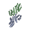
| ||||||||||
|---|---|---|---|---|---|---|---|---|---|---|---|
| 1 | 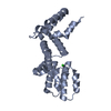
| ||||||||||
| 2 | 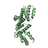
| ||||||||||
| Unit cell |
|
-Components
| #1: Protein | Mass: 26258.492 Da / Num. of mol.: 2 Source method: isolated from a genetically manipulated source Source: (gene. exp.) Haemophilus influenzae (bacteria) / Strain: ATCC 51907 / DSM 11121 / KW20 / Rd / Gene: lpoA, HI_1655 / Plasmid: pETBlue2Details (production host): T7 promoter and C-terminal His6 tag Production host: #2: Chemical |   Mass: 35.453 Da / Num. of mol.: 2 / Source method: obtained synthetically / Formula: Cl Mass: 35.453 Da / Num. of mol.: 2 / Source method: obtained synthetically / Formula: Cl#3: Water | ChemComp-HOH / |  Mass: 18.015 Da / Num. of mol.: 683 / Source method: isolated from a natural source / Formula: H2O Mass: 18.015 Da / Num. of mol.: 683 / Source method: isolated from a natural source / Formula: H2O |
|---|
-Experimental details
-Experiment
| Experiment | Method: X-RAY DIFFRACTION / Number of used crystals: 1 |
|---|
- Sample preparation
| Crystal | Density Matthews: 2.31 Å3/Da / Density % sol: 46.74 % |
|---|---|
| Crystal grow | Temperature: 295 K / Method: vapor diffusion, hanging drop Details: Protein was 10 mg/ml in 0.1% beta-mercaptoethanol, 1 mM EDTA, 0.1 mM benzamidine, 20 mM Tris-HCl pH 8.0. Precipitant contained 20% polyethylene glycol 10,000, 0.1 M HEPES pH 7.5. 2 ul ...Details: Protein was 10 mg/ml in 0.1% beta-mercaptoethanol, 1 mM EDTA, 0.1 mM benzamidine, 20 mM Tris-HCl pH 8.0. Precipitant contained 20% polyethylene glycol 10,000, 0.1 M HEPES pH 7.5. 2 ul protein mixed with 2 ul precipitant on silated cover slip and equilibrated over a reservoir containing 1 ml precipitant. PH range: 7.5-8.0 |
-Data collection
| Diffraction | Mean temperature: 140 K | |||||||||||||||||||||||||||||||||||||||||||||||||||||||||||||||||||||||||||||
|---|---|---|---|---|---|---|---|---|---|---|---|---|---|---|---|---|---|---|---|---|---|---|---|---|---|---|---|---|---|---|---|---|---|---|---|---|---|---|---|---|---|---|---|---|---|---|---|---|---|---|---|---|---|---|---|---|---|---|---|---|---|---|---|---|---|---|---|---|---|---|---|---|---|---|---|---|---|---|
| Diffraction source | Source: SYNCHROTRON / Site: APS  / Beamline: 5ID-B / Wavelength: 0.9793 Å / Beamline: 5ID-B / Wavelength: 0.9793 Å | |||||||||||||||||||||||||||||||||||||||||||||||||||||||||||||||||||||||||||||
| Detector | Type: RAYONIX MX225-HS / Detector: CCD / Date: Nov 12, 2003 | |||||||||||||||||||||||||||||||||||||||||||||||||||||||||||||||||||||||||||||
| Radiation | Protocol: SINGLE WAVELENGTH / Monochromatic (M) / Laue (L): M / Scattering type: x-ray | |||||||||||||||||||||||||||||||||||||||||||||||||||||||||||||||||||||||||||||
| Radiation wavelength | Wavelength: 0.9793 Å / Relative weight: 1 | |||||||||||||||||||||||||||||||||||||||||||||||||||||||||||||||||||||||||||||
| Reflection | Resolution: 1.35→34.5 Å / Num. obs: 91763 / % possible obs: 90.3 % / Redundancy: 3.28 % / Biso Wilson estimate: 15.58 Å2 / Rmerge(I) obs: 0.047 / Rrim(I) all: 0.047 / Χ2: 0.97 / Net I/σ(I): 13.1 / Num. measured all: 303668 / Scaling rejects: 2279 | |||||||||||||||||||||||||||||||||||||||||||||||||||||||||||||||||||||||||||||
| Reflection shell | Diffraction-ID: 1
|
-Phasing
| Phasing | Method: molecular replacement | |||||||||
|---|---|---|---|---|---|---|---|---|---|---|
| Phasing MR |
|
- Processing
| Software |
| |||||||||||||||||||||||||||||||||||||||||||||||||||||||||||||||||||||||||||||||||||||||||||||||||||||||||||||||||||||||||||||||||||||||||||||||||||||||||||||||||||||||||||||||||||||||||||||||||||||||||||||||||||||||||
|---|---|---|---|---|---|---|---|---|---|---|---|---|---|---|---|---|---|---|---|---|---|---|---|---|---|---|---|---|---|---|---|---|---|---|---|---|---|---|---|---|---|---|---|---|---|---|---|---|---|---|---|---|---|---|---|---|---|---|---|---|---|---|---|---|---|---|---|---|---|---|---|---|---|---|---|---|---|---|---|---|---|---|---|---|---|---|---|---|---|---|---|---|---|---|---|---|---|---|---|---|---|---|---|---|---|---|---|---|---|---|---|---|---|---|---|---|---|---|---|---|---|---|---|---|---|---|---|---|---|---|---|---|---|---|---|---|---|---|---|---|---|---|---|---|---|---|---|---|---|---|---|---|---|---|---|---|---|---|---|---|---|---|---|---|---|---|---|---|---|---|---|---|---|---|---|---|---|---|---|---|---|---|---|---|---|---|---|---|---|---|---|---|---|---|---|---|---|---|---|---|---|---|---|---|---|---|---|---|---|---|---|---|---|---|---|---|---|---|
| Refinement | Method to determine structure: MOLECULAR REPLACEMENT Starting model: 4P29 Resolution: 1.35→34.44 Å / SU ML: 0.1352 / Cross valid method: THROUGHOUT / σ(F): 1.36 / Phase error: 17.9643
| |||||||||||||||||||||||||||||||||||||||||||||||||||||||||||||||||||||||||||||||||||||||||||||||||||||||||||||||||||||||||||||||||||||||||||||||||||||||||||||||||||||||||||||||||||||||||||||||||||||||||||||||||||||||||
| Solvent computation | Shrinkage radii: 0.9 Å / VDW probe radii: 1.11 Å | |||||||||||||||||||||||||||||||||||||||||||||||||||||||||||||||||||||||||||||||||||||||||||||||||||||||||||||||||||||||||||||||||||||||||||||||||||||||||||||||||||||||||||||||||||||||||||||||||||||||||||||||||||||||||
| Displacement parameters | Biso mean: 25.85 Å2 | |||||||||||||||||||||||||||||||||||||||||||||||||||||||||||||||||||||||||||||||||||||||||||||||||||||||||||||||||||||||||||||||||||||||||||||||||||||||||||||||||||||||||||||||||||||||||||||||||||||||||||||||||||||||||
| Refinement step | Cycle: LAST / Resolution: 1.35→34.44 Å
| |||||||||||||||||||||||||||||||||||||||||||||||||||||||||||||||||||||||||||||||||||||||||||||||||||||||||||||||||||||||||||||||||||||||||||||||||||||||||||||||||||||||||||||||||||||||||||||||||||||||||||||||||||||||||
| Refine LS restraints |
| |||||||||||||||||||||||||||||||||||||||||||||||||||||||||||||||||||||||||||||||||||||||||||||||||||||||||||||||||||||||||||||||||||||||||||||||||||||||||||||||||||||||||||||||||||||||||||||||||||||||||||||||||||||||||
| LS refinement shell |
|
