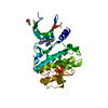
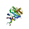
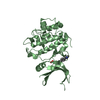
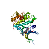
- PDB-6d8x: PPAR gamma LBD complexed with the agonist GW1929 -
+
Open data
ID or keywords:
Loading...
-
Basic information
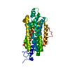
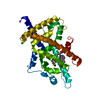
Entry
Database: PDB / ID: 6d8x
Title
PPAR gamma LBD complexed with the agonist GW1929
Components
Peroxisome proliferator-activated receptor gamma
Keywords
dna binding protein/AGONIST / Nuclear receptor / Transcription factor / Super agonist / Ligand binding domain / DNA BINDING PROTEIN / dna binding protein-AGONIST complex
Function / homology
Function and homology information
prostaglandin receptor activity / negative regulation of receptor signaling pathway via STAT / MECP2 regulates transcription factors / beige fat cell differentiation / negative regulation of vascular endothelial cell proliferation / negative regulation of extracellular matrix assembly / negative regulation of connective tissue replacement involved in inflammatory response wound healing / positive regulation of cholesterol transport / negative regulation of cellular response to transforming growth factor beta stimulus / arachidonate binding ...prostaglandin receptor activity / negative regulation of receptor signaling pathway via STAT / MECP2 regulates transcription factors / beige fat cell differentiation / negative regulation of vascular endothelial cell proliferation / negative regulation of extracellular matrix assembly / negative regulation of connective tissue replacement involved in inflammatory response wound healing / positive regulation of cholesterol transport / negative regulation of cellular response to transforming growth factor beta stimulus / arachidonate binding / positive regulation of adiponectin secretion / DNA binding domain binding / negative regulation of cardiac muscle hypertrophy in response to stress / positive regulation of vascular associated smooth muscle cell apoptotic process / positive regulation of lipid metabolic process / positive regulation of fatty acid metabolic process / STAT family protein binding / WW domain binding / negative regulation of type II interferon-mediated signaling pathway / LBD domain binding / negative regulation of cholesterol storage / response to lipid / positive regulation of lipoprotein transport / negative regulation of SMAD protein signal transduction / lipid homeostasis / E-box binding / R-SMAD binding / negative regulation of blood vessel endothelial cell migration / white fat cell differentiation / alpha-actinin binding / negative regulation of vascular associated smooth muscle cell proliferation / negative regulation of macrophage derived foam cell differentiation / negative regulation of lipid storage / positive regulation of cholesterol efflux / negative regulation of BMP signaling pathway / monocyte differentiation / cell fate commitment / cellular response to low-density lipoprotein particle stimulus / long-chain fatty acid transport / BMP signaling pathway / negative regulation of mitochondrial fission / negative regulation of osteoblast differentiation / positive regulation of fat cell differentiation / nuclear retinoid X receptor binding / fat cell differentiation / Transcriptional regulation of brown and beige adipocyte differentiation by EBF2 / retinoic acid receptor signaling pathway / intracellular receptor signaling pathway / negative regulation of MAPK cascade / peptide binding / peroxisome proliferator activated receptor signaling pathway / cell maturation / epithelial cell differentiation / hormone-mediated signaling pathway / regulation of cellular response to insulin stimulus / positive regulation of adipose tissue development / response to nutrient / negative regulation of miRNA transcription / brown fat cell differentiation / negative regulation of angiogenesis / placenta development / Regulation of PTEN gene transcription / transcription coregulator binding / SUMOylation of intracellular receptors / positive regulation of apoptotic signaling pathway / negative regulation of smooth muscle cell proliferation / negative regulation of transforming growth factor beta receptor signaling pathway / PPARA activates gene expression / fatty acid metabolic process / Nuclear Receptor transcription pathway / Transcriptional regulation of white adipocyte differentiation / regulation of circadian rhythm / positive regulation of miRNA transcription / mRNA transcription by RNA polymerase II / DNA-binding transcription repressor activity, RNA polymerase II-specific / nuclear receptor activity / negative regulation of inflammatory response / regulation of blood pressure / RNA polymerase II transcription regulator complex / cellular response to insulin stimulus / rhythmic process / glucose homeostasis / MLL4 and MLL3 complexes regulate expression of PPARG target genes in adipogenesis and hepatic steatosis / double-stranded DNA binding / DNA-binding transcription activator activity, RNA polymerase II-specific / cellular response to hypoxia / DNA-binding transcription factor binding / sequence-specific DNA binding / nucleic acid binding / DNA-binding transcription factor activity, RNA polymerase II-specific / cell differentiation / signaling receptor complex / transcription cis-regulatory region binding / RNA polymerase II cis-regulatory region sequence-specific DNA binding / DNA-binding transcription factor activity / negative regulation of gene expression / innate immune response / negative regulation of DNA-templated transcription / chromatin binding / positive regulation of gene expression Similarity search - Function
In the structure databanks used in Yorodumi, some data are registered as the other names, "COVID-19 virus" and "2019-nCoV". Here are the details of the virus and the list of structure data.
Jan 31, 2019. EMDB accession codes are about to change! (news from PDBe EMDB page)
EMDB accession codes are about to change! (news from PDBe EMDB page)
The allocation of 4 digits for EMDB accession codes will soon come to an end. Whilst these codes will remain in use, new EMDB accession codes will include an additional digit and will expand incrementally as the available range of codes is exhausted. The current 4-digit format prefixed with “EMD-” (i.e. EMD-XXXX) will advance to a 5-digit format (i.e. EMD-XXXXX), and so on. It is currently estimated that the 4-digit codes will be depleted around Spring 2019, at which point the 5-digit format will come into force.
The EM Navigator/Yorodumi systems omit the EMD- prefix.
Related info.:Q: What is EMD? / ID/Accession-code notation in Yorodumi/EM Navigator
Yorodumi is a browser for structure data from EMDB, PDB, SASBDB, etc.
This page is also the successor to EM Navigator detail page, and also detail information page/front-end page for Omokage search.
The word "yorodu" (or yorozu) is an old Japanese word meaning "ten thousand". "mi" (miru) is to see.
Related info.:EMDB / PDB / SASBDB / Comparison of 3 databanks / Yorodumi Search / Aug 31, 2016. New EM Navigator & Yorodumi / Yorodumi Papers / Jmol/JSmol / Function and homology information / Changes in new EM Navigator and Yorodumi
 Movie
Movie Controller


 Open data
 Basic information
 Components
 Keywords
 Function and homology information
 Homo sapiens (human)
 X-RAY DIFFRACTION /
  Authors
 United States, 2items
  Citation
 Structure visualization
 Downloads & links
 Other downloads


 PDBj
PDBj


 Assembly

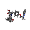


 Mass: 495.569 Da / Num. of mol.: 1 / Source method: obtained synthetically / Formula: C30H29N3O4
 Mass: 189.100 Da / Num. of mol.: 1 / Source method: obtained synthetically / Formula: C6H5O7
 Mass: 22.990 Da / Num. of mol.: 3 / Source method: obtained synthetically / Formula: Na
 Mass: 92.094 Da / Num. of mol.: 1 / Source method: obtained synthetically / Formula: C3H8O3
 Sample preparation
 Processing