ムービー
ムービー コントローラー


+ データを開く

- 基本情報
基本情報
| 登録情報 | データベース: PDB / ID: 6d8c | |||||||||
|---|---|---|---|---|---|---|---|---|---|---|
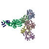
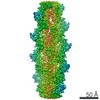
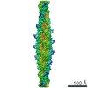
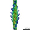
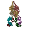
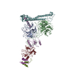
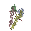
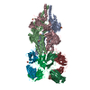
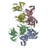
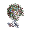
| タイトル | Cryo-EM structure of FLNaABD E254K bound to phalloidin-stabilized F-actin | |||||||||
 要素 要素 |
| |||||||||
 キーワード キーワード | STRUCTURAL PROTEIN / Actin-binding domain / Actin crosslinker | |||||||||
| 機能・相同性 |  機能・相同性情報 機能・相同性情報regulation of membrane repolarization during atrial cardiac muscle cell action potential / regulation of membrane repolarization during cardiac muscle cell action potential / establishment of Sertoli cell barrier / Myb complex / glycoprotein Ib-IX-V complex / adenylate cyclase-inhibiting dopamine receptor signaling pathway / formation of radial glial scaffolds / positive regulation of integrin-mediated signaling pathway / actin crosslink formation / blood coagulation, intrinsic pathway ...regulation of membrane repolarization during atrial cardiac muscle cell action potential / regulation of membrane repolarization during cardiac muscle cell action potential / establishment of Sertoli cell barrier / Myb complex / glycoprotein Ib-IX-V complex / adenylate cyclase-inhibiting dopamine receptor signaling pathway / formation of radial glial scaffolds / positive regulation of integrin-mediated signaling pathway / actin crosslink formation / blood coagulation, intrinsic pathway / tubulin deacetylation / OAS antiviral response / positive regulation of actin filament bundle assembly / Striated Muscle Contraction / positive regulation of neuron migration / protein localization to bicellular tight junction / Fc-gamma receptor I complex binding / Cell-extracellular matrix interactions / apical dendrite / positive regulation of potassium ion transmembrane transport / positive regulation of neural precursor cell proliferation / positive regulation of platelet activation / protein localization to cell surface / wound healing, spreading of cells / podosome / negative regulation of transcription by RNA polymerase I / megakaryocyte development / GP1b-IX-V activation signalling / SMAD binding / receptor clustering / cortical cytoskeleton / RHO GTPases activate PAKs / semaphorin-plexin signaling pathway / striated muscle thin filament / skeletal muscle thin filament assembly / cilium assembly / mitotic spindle assembly / potassium channel regulator activity / negative regulation of DNA-binding transcription factor activity / skeletal muscle fiber development / stress fiber / release of sequestered calcium ion into cytosol / positive regulation of substrate adhesion-dependent cell spreading / protein sequestering activity / regulation of cell migration / dendritic shaft / protein localization to plasma membrane / actin filament / establishment of protein localization / mRNA transcription by RNA polymerase II / G protein-coupled receptor binding / cerebral cortex development / negative regulation of protein catabolic process / positive regulation of protein import into nucleus / small GTPase binding / 加水分解酵素; 酸無水物に作用; 酸無水物に作用・細胞または細胞小器官の運動に関与 / platelet aggregation / kinase binding / Z disc / actin filament binding / cell-cell junction / Platelet degranulation / actin cytoskeleton / growth cone / toxin activity / GTPase binding / actin cytoskeleton organization / perikaryon / DNA-binding transcription factor binding / transmembrane transporter binding / hydrolase activity / positive regulation of canonical NF-kappaB signal transduction / postsynapse / protein stabilization / cadherin binding / focal adhesion / negative regulation of apoptotic process / nucleolus / perinuclear region of cytoplasm / glutamatergic synapse / protein homodimerization activity / RNA binding / extracellular exosome / extracellular region / ATP binding / nucleus / membrane / plasma membrane / cytosol / cytoplasm 類似検索 - 分子機能 | |||||||||
| 生物種 |  Homo sapiens (ヒト) Homo sapiens (ヒト)  Amanita phalloides (タマゴテングタケ) Amanita phalloides (タマゴテングタケ) | |||||||||
| 手法 | 電子顕微鏡法 / らせん対称体再構成法 / クライオ電子顕微鏡法 / 解像度: 3.54 Å | |||||||||
 データ登録者 データ登録者 | Iwamoto, D.V. / Huehn, A.R. / Simon, B. / Huet-Calderwood, C. / Baldassarre, M. / Sindelar, C.V. / Calderwood, D.A. | |||||||||
 引用 引用 | ジャーナル: Nat Struct Mol Biol / 年: 2018 タイトル: Structural basis of the filamin A actin-binding domain interaction with F-actin. 著者: Daniel V Iwamoto / Andrew Huehn / Bertrand Simon / Clotilde Huet-Calderwood / Massimiliano Baldassarre / Charles V Sindelar / David A Calderwood /   要旨: Actin-cross-linking proteins assemble actin filaments into higher-order structures essential for orchestrating cell shape, adhesion, and motility. Missense mutations in the tandem calponin homology ...Actin-cross-linking proteins assemble actin filaments into higher-order structures essential for orchestrating cell shape, adhesion, and motility. Missense mutations in the tandem calponin homology domains of their actin-binding domains (ABDs) underlie numerous genetic diseases, but a molecular understanding of these pathologies is hampered by the lack of high-resolution structures of any actin-cross-linking protein bound to F-actin. Here, taking advantage of a high-affinity, disease-associated mutant of the human filamin A (FLNa) ABD, we combine cryo-electron microscopy and functional studies to reveal at near-atomic resolution how the first calponin homology domain (CH1) and residues immediately N-terminal to it engage actin. We further show that reorientation of CH2 relative to CH1 is required to avoid clashes with actin and to expose F-actin-binding residues on CH1. Our data explain localization of disease-associated loss-of-function mutations to FLNaCH1 and gain-of-function mutations to the regulatory FLNaCH2. Sequence conservation argues that this provides a general model for ABD-F-actin binding. | |||||||||
| 履歴 |
|
- 構造の表示
構造の表示
| ムービー |
ムービービューア |
|---|---|
| 構造ビューア | 分子: MolmilJmol/JSmol |
- ダウンロードとリンク
-ダウンロード
| PDBx/mmCIF形式 | 6d8c.cif.gz | 854.1 KB | 表示 | PDBx/mmCIF形式 |
|---|---|---|---|---|
| PDB形式 | pdb6d8c.ent.gz | 711.6 KB | 表示 | PDB形式 |
| PDBx/mmJSON形式 | 6d8c.json.gz | ツリー表示 | PDBx/mmJSON形式 | |
| その他 |  その他のダウンロード その他のダウンロード |
-検証レポート
| 文書・要旨 | 6d8c_validation.pdf.gz | 1.4 MB | 表示 | wwPDB検証レポート |
|---|---|---|---|---|
| 文書・詳細版 | 6d8c_full_validation.pdf.gz | 1.4 MB | 表示 | |
| XML形式データ | 6d8c_validation.xml.gz | 76.6 KB | 表示 | |
| CIF形式データ | 6d8c_validation.cif.gz | 114.3 KB | 表示 | |
| アーカイブディレクトリ | https://data.pdbj.org/pub/pdb/validation_reports/d8/6d8cftp://data.pdbj.org/pub/pdb/validation_reports/d8/6d8c | HTTPS FTP |
-関連構造データ

-リンク
 PDBj
PDBj


- 集合体
集合体
| 登録構造単位 | 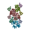
|
|---|---|
| 1 |
|
-要素
| #1: タンパク質 | 分子量: 34476.250 Da / 分子数: 5 / 断片: UNP residues 1-278 / 変異: E254K / 由来タイプ: 組換発現 / 由来: (組換発現) Homo sapiens (ヒト) / 遺伝子: FLNA, FLN, FLN1 / 発現宿主:  #2: タンパク質 | 分子量: 41862.613 Da / 分子数: 5 / 由来タイプ: 天然 / 由来: (天然) #3: タンパク質・ペプチド |   タイプ: Cyclic peptide / クラス: 毒素 / 分子量: 808.899 Da / 分子数: 3 / 由来タイプ: 天然 タイプ: Cyclic peptide / クラス: 毒素 / 分子量: 808.899 Da / 分子数: 3 / 由来タイプ: 天然由来: (天然) Amanita phalloides (タマゴテングタケ)参照: Phalloidin, UniProt: P0CU65*PLUS #4: 化合物 | ChemComp-ADP /   分子量: 427.201 Da / 分子数: 5 / 由来タイプ: 合成 / 式: C10H15N5O10P2 / コメント: ADP, エネルギー貯蔵分子*YM 分子量: 427.201 Da / 分子数: 5 / 由来タイプ: 合成 / 式: C10H15N5O10P2 / コメント: ADP, エネルギー貯蔵分子*YM#5: 化合物 | ChemComp-MG /   分子量: 24.305 Da / 分子数: 5 / 由来タイプ: 合成 / 式: Mg 分子量: 24.305 Da / 分子数: 5 / 由来タイプ: 合成 / 式: Mg |
|---|
-実験情報
-実験
| 実験 | 手法: 電子顕微鏡法 |
|---|---|
| EM実験 | 試料の集合状態: FILAMENT / 3次元再構成法: らせん対称体再構成法 |
- 試料調製
試料調製
| 構成要素 | 名称: Helical complex of FLNaABD E254K bound to phalloidin-stabilized F-actin タイプ: COMPLEX / Entity ID: #1-#3 / 由来: MULTIPLE SOURCES |
|---|---|
| 由来(天然) | 生物種: Homo sapiens (ヒト) |
| 由来(組換発現) | 生物種: |
| 緩衝液 | pH: 8 |
| 試料 | 包埋: NO / シャドウイング: NO / 染色: NO / 凍結: YES |
| 急速凍結 | 装置: HOMEMADE PLUNGER / 凍結剤: ETHANE |
- 電子顕微鏡撮影
| 実験機器 |  モデル: Titan Krios / 画像提供: FEI Company |
|---|---|
| 顕微鏡 | モデル: FEI TITAN KRIOS |
| 電子銃 | 電子線源:  FIELD EMISSION GUN / 加速電圧: 300 kV / 照射モード: SPOT SCAN FIELD EMISSION GUN / 加速電圧: 300 kV / 照射モード: SPOT SCAN |
| 電子レンズ | モード: BRIGHT FIELD / 倍率(公称値): 105000 X / 倍率(補正後): 37500 X / 最大 デフォーカス(公称値): 2900 nm / 最小 デフォーカス(公称値): 1000 nm / Cs: 2.7 mm / C2レンズ絞り径: 50 µm / アライメント法: COMA FREE |
| 試料ホルダ | 試料ホルダーモデル: FEI TITAN KRIOS AUTOGRID HOLDER |
| 撮影 | 平均露光時間: 12 sec. / 電子線照射量: 50 e/Å2 / 検出モード: SUPER-RESOLUTION フィルム・検出器のモデル: GATAN K2 SUMMIT (4k x 4k) 撮影したグリッド数: 2 / 実像数: 2140 詳細: Micrographs from only one of the grids were used in the reconstruction. From that grid, only micrographs where Gctf detected signal at resolutions better than 4A were used in the reconstruction (~15%). |
| 画像スキャン | 動画フレーム数/画像: 40 / 利用したフレーム数/画像: 1-40 |
- 解析
解析
| EMソフトウェア |
| ||||||||||||||||||||||||||||
|---|---|---|---|---|---|---|---|---|---|---|---|---|---|---|---|---|---|---|---|---|---|---|---|---|---|---|---|---|---|
| CTF補正 | タイプ: PHASE FLIPPING AND AMPLITUDE CORRECTION | ||||||||||||||||||||||||||||
| らせん対称 | 回転角度/サブユニット: -166.73 ° / 軸方向距離/サブユニット: 27.54 Å / らせん対称軸の対称性: C1 | ||||||||||||||||||||||||||||
| 粒子像の選択 | 選択した粒子像数: 67000 | ||||||||||||||||||||||||||||
| 3次元再構成 | 解像度: 3.54 Å / 解像度の算出法: FSC 0.143 CUT-OFF / 粒子像の数: 67000 / 対称性のタイプ: HELICAL | ||||||||||||||||||||||||||||
| 原子モデル構築 | プロトコル: RIGID BODY FIT | ||||||||||||||||||||||||||||
| 原子モデル構築 | 3D fitting-ID: 1 / Source name: PDB / タイプ: experimental model
|