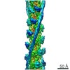
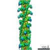
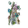
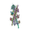
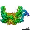
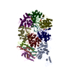
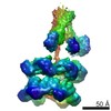
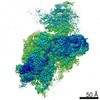
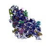
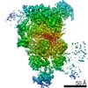
登録情報 データベース : PDB / ID : 6cxiタイトル Cardiac thin filament decorated with C0C1 fragment of cardiac myosin binding protein C mode 1 (Myosin-binding protein C, cardiac-type) x 2 Actin, cytoplasmic 2 Tropomyosin キーワード / 機能・相同性 分子機能 ドメイン・相同性 構成要素
/ / / / / / / / / / / / / / / / / / / / / / / / / / / / / / / / / / / / / / / / / / / / / / / / / / / / / / / / / / / / / / / / / / / / / / / / / / / / / / / / / / / / / / / / / / / / / / / / / / / / / / / / / / / / / / / / / / / / / / / / / / / / / / / / / / / / / / / / / / / / / / 生物種 Homo sapiens (ヒト)手法 / / / 解像度 : 11 Å データ登録者 Galkin, V.E. / Schroeder, G.F. 資金援助 組織 認可番号 国 American Heart Association 560851
ジャーナル : Structure / 年 : 2018タイトル : N-Terminal Domains of Cardiac Myosin Binding Protein C Cooperatively Activate the Thin Filament.著者 : Cristina Risi / Betty Belknap / Eva Forgacs-Lonart / Samantha P Harris / Gunnar F Schröder / Howard D White / Vitold E Galkin / 要旨 : Muscle contraction relies on interaction between myosin-based thick filaments and actin-based thin filaments. Myosin binding protein C (MyBP-C) is a key regulator of actomyosin interactions. Recent ... Muscle contraction relies on interaction between myosin-based thick filaments and actin-based thin filaments. Myosin binding protein C (MyBP-C) is a key regulator of actomyosin interactions. Recent studies established that the N'-terminal domains (NTDs) of MyBP-C can either activate or inhibit thin filaments, but the mechanism of their collective action is poorly understood. Cardiac MyBP-C (cMyBP-C) harbors an extra NTD, which is absent in skeletal isoforms of MyBP-C, and its role in regulation of cardiac contraction is unknown. Here we show that the first two domains of human cMyPB-C (i.e., C0 and C1) cooperate to activate the thin filament. We demonstrate that C1 interacts with tropomyosin via a positively charged loop and that this interaction, stabilized by the C0 domain, is required for thin filament activation by cMyBP-C. Our data reveal a mechanism by which cMyBP-C can modulate cardiac contraction and demonstrate a function of the C0 domain. 履歴 登録 2018年4月3日 登録サイト / 処理サイト 改定 1.0 2018年10月10日 Provider / タイプ 改定 1.1 2018年12月19日 Group / Database references / Structure summaryカテゴリ / entityItem / _citation.page_first / _entity.formula_weight改定 1.2 2024年3月13日 Group / Database references / カテゴリ / chem_comp_bond / database_2Item / _database_2.pdbx_database_accession
すべて表示 表示を減らす
 ムービー
ムービー コントローラー


 データを開く
 基本情報
基本情報 要素
要素 キーワード
キーワード 機能・相同性情報
 Homo sapiens (ヒト)
 データ登録者
 米国, 1件
  引用
引用
 構造の表示
構造の表示 ダウンロードとリンク
 その他のダウンロード
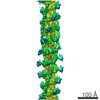


 PDBj
PDBj


 集合体
集合体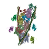

 試料調製
試料調製 電子顕微鏡撮影
 FIELD EMISSION GUN / 加速電圧: 300 kV / 照射モード: FLOOD BEAM
 解析
解析