 Movie
Movie Controller

[English] 日本語
 Yorodumi
- PDB-6swd: IC2 body model of cryo-EM structure of a full archaeal ribosomal ... -
+ Open data

- Basic information
| Entry | Database: PDB / ID: 6swd | |||||||||||||||||||||||||||||||||||||||||||||||||||||||||||||||||||||||||||
|---|---|---|---|---|---|---|---|---|---|---|---|---|---|---|---|---|---|---|---|---|---|---|---|---|---|---|---|---|---|---|---|---|---|---|---|---|---|---|---|---|---|---|---|---|---|---|---|---|---|---|---|---|---|---|---|---|---|---|---|---|---|---|---|---|---|---|---|---|---|---|---|---|---|---|---|---|
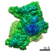
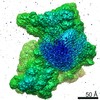
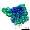
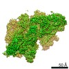
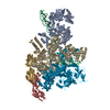
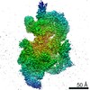
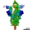
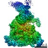
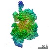
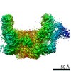
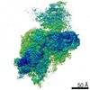
| Title | IC2 body model of cryo-EM structure of a full archaeal ribosomal translation initiation complex devoid of aIF1 in P. abyssi | |||||||||||||||||||||||||||||||||||||||||||||||||||||||||||||||||||||||||||
 Components Components |
| |||||||||||||||||||||||||||||||||||||||||||||||||||||||||||||||||||||||||||
 Keywords Keywords | RIBOSOME / Translation initiation / cryo-EM / tRNA / evolution / archaea / rRNA modifications | |||||||||||||||||||||||||||||||||||||||||||||||||||||||||||||||||||||||||||
| Function / homology |  Function and homology information Function and homology informationtranslation initiation factor activity / ribosomal small subunit biogenesis / small ribosomal subunit / small ribosomal subunit rRNA binding / cytosolic small ribosomal subunit / rRNA binding / structural constituent of ribosome / ribosome / translation / ribonucleoprotein complex ...translation initiation factor activity / ribosomal small subunit biogenesis / small ribosomal subunit / small ribosomal subunit rRNA binding / cytosolic small ribosomal subunit / rRNA binding / structural constituent of ribosome / ribosome / translation / ribonucleoprotein complex / RNA binding / zinc ion binding Similarity search - Function | |||||||||||||||||||||||||||||||||||||||||||||||||||||||||||||||||||||||||||
| Biological species |   Pyrococcus abyssi (archaea)Pyrococcus abyssi GE5 (archaea) Pyrococcus abyssi (archaea)Pyrococcus abyssi GE5 (archaea) | |||||||||||||||||||||||||||||||||||||||||||||||||||||||||||||||||||||||||||
| Method | ELECTRON MICROSCOPY / single particle reconstruction / cryo EM / Resolution: 3.2 Å | |||||||||||||||||||||||||||||||||||||||||||||||||||||||||||||||||||||||||||
 Authors Authors | Coureux, P.-D. / Mechulam, Y. / Schmitt, E. | |||||||||||||||||||||||||||||||||||||||||||||||||||||||||||||||||||||||||||
| Funding support |  France, 1items France, 1items
| |||||||||||||||||||||||||||||||||||||||||||||||||||||||||||||||||||||||||||
 Citation Citation | Journal: Commun Biol / Year: 2020 Title: Cryo-EM study of an archaeal 30S initiation complex gives insights into evolution of translation initiation. Authors: Pierre-Damien Coureux / Christine Lazennec-Schurdevin / Sophie Bourcier / Yves Mechulam / Emmanuelle Schmitt / Abstract: Archaeal translation initiation occurs within a macromolecular complex containing the small ribosomal subunit (30S) bound to mRNA, initiation factors aIF1, aIF1A and the ternary complex aIF2:GDPNP: ...Archaeal translation initiation occurs within a macromolecular complex containing the small ribosomal subunit (30S) bound to mRNA, initiation factors aIF1, aIF1A and the ternary complex aIF2:GDPNP:Met-tRNA. Here, we determine the cryo-EM structure of a 30S:mRNA:aIF1A:aIF2:GTP:Met-tRNA complex from Pyrococcus abyssi at 3.2 Å resolution. It highlights archaeal features in ribosomal proteins and rRNA modifications. We find an aS21 protein, at the location of eS21 in eukaryotic ribosomes. Moreover, we identify an N-terminal extension of archaeal eL41 contacting the P site. We characterize 34 N-acetylcytidines distributed throughout 16S rRNA, likely contributing to hyperthermostability. Without aIF1, the 30S head is stabilized and initiator tRNA is tightly bound to the P site. A network of interactions involving tRNA, mRNA, rRNA modified nucleotides and C-terminal tails of uS9, uS13 and uS19 is observed. Universal features and domain-specific idiosyncrasies of translation initiation are discussed in light of ribosomal structures from representatives of each domain of life. | |||||||||||||||||||||||||||||||||||||||||||||||||||||||||||||||||||||||||||
| History |
|
- Structure visualization
| Movie |
Movie viewer |
|---|---|
| Structure viewer | Molecule: MolmilJmol/JSmol |
- Downloads & links
-Download
| PDBx/mmCIF format | 6swd.cif.gz | 944 KB | Display | PDBx/mmCIF format |
|---|---|---|---|---|
| PDB format | pdb6swd.ent.gz | 728.3 KB | Display | PDB format |
| PDBx/mmJSON format | 6swd.json.gz | Tree view | PDBx/mmJSON format | |
| Others |  Other downloads Other downloads |
-Validation report
| Arichive directory | https://data.pdbj.org/pub/pdb/validation_reports/sw/6swdftp://data.pdbj.org/pub/pdb/validation_reports/sw/6swd | HTTPS FTP |
|---|
-Related structure data
| Related structure data | 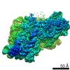 10323MC 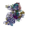 6sw9C  6swcC 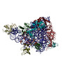 6sweC M: map data used to model this data C: citing same article ( |
|---|---|
| Similar structure data |
-Links
 PDBj
PDBj


- Assembly
| Deposited unit | 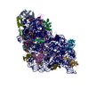
|
|---|---|
| 1 |
|
-Components
-RNA chain , 2 types, 2 molecules 25
| #1: RNA chain | Mass: 340430.125 Da / Num. of mol.: 1 / Source method: isolated from a natural source / Source: (natural) Pyrococcus abyssi GE5 (archaea) |
|---|---|
| #18: RNA chain | Mass: 6448.871 Da / Num. of mol.: 1 / Source method: obtained synthetically / Source: (synth.) Pyrococcus abyssi GE5 (archaea) |
-30S ribosomal protein ... , 15 types, 15 molecules ABDEFGIJMNQRVW0
| #2: Protein | Mass: 22888.068 Da / Num. of mol.: 1 / Source method: isolated from a natural source Source: (natural) Pyrococcus abyssi (strain GE5 / Orsay) (archaea)Strain: GE5 / Orsay / References: UniProt: Q9V2K7 |
|---|---|
| #3: Protein | Mass: 23026.865 Da / Num. of mol.: 1 / Source method: isolated from a natural source Source: (natural) Pyrococcus abyssi (strain GE5 / Orsay) (archaea)Strain: GE5 / Orsay / References: UniProt: Q9V191 |
| #5: Protein | Mass: 21381.910 Da / Num. of mol.: 1 / Source method: isolated from a natural source Source: (natural) Pyrococcus abyssi (strain GE5 / Orsay) (archaea)Strain: GE5 / Orsay / References: UniProt: P61992 |
| #6: Protein | Mass: 28246.119 Da / Num. of mol.: 1 / Source method: isolated from a natural source Source: (natural) Pyrococcus abyssi (strain GE5 / Orsay) (archaea)Strain: GE5 / Orsay / References: UniProt: Q9V1U8 |
| #7: Protein | Mass: 26523.904 Da / Num. of mol.: 1 / Source method: isolated from a natural source Source: (natural) Pyrococcus abyssi (strain GE5 / Orsay) (archaea)Strain: GE5 / Orsay / References: UniProt: Q9V1V5 |
| #8: Protein | Mass: 14029.461 Da / Num. of mol.: 1 / Source method: isolated from a natural source Source: (natural) Pyrococcus abyssi (strain GE5 / Orsay) (archaea)Strain: GE5 / Orsay / References: UniProt: Q9UYS3 |
| #9: Protein | Mass: 14772.246 Da / Num. of mol.: 1 / Source method: isolated from a natural source Source: (natural) Pyrococcus abyssi (strain GE5 / Orsay) (archaea)Strain: GE5 / Orsay / References: UniProt: Q9V1V0 |
| #10: Protein | Mass: 14293.733 Da / Num. of mol.: 1 / Source method: isolated from a natural source Source: (natural) Pyrococcus abyssi (strain GE5 / Orsay) (archaea)Strain: GE5 / Orsay / References: UniProt: Q9UZL4 |
| #11: Protein | Mass: 14772.950 Da / Num. of mol.: 1 / Source method: isolated from a natural source Source: (natural) Pyrococcus abyssi (strain GE5 / Orsay) (archaea)Strain: GE5 / Orsay / References: UniProt: P62010 |
| #12: Protein | Mass: 16477.621 Da / Num. of mol.: 1 / Source method: isolated from a natural source Source: (natural) Pyrococcus abyssi (strain GE5 / Orsay) (archaea)Strain: GE5 / Orsay / References: UniProt: P61994 |
| #13: Protein | Mass: 18697.143 Da / Num. of mol.: 1 / Source method: isolated from a natural source Source: (natural) Pyrococcus abyssi (strain GE5 / Orsay) (archaea)Strain: GE5 / Orsay / References: UniProt: Q9V2K9 |
| #14: Protein | Mass: 13364.604 Da / Num. of mol.: 1 / Source method: isolated from a natural source Source: (natural) Pyrococcus abyssi (strain GE5 / Orsay) (archaea)Strain: GE5 / Orsay / References: UniProt: Q9V1U5 |
| #15: Protein | Mass: 11798.637 Da / Num. of mol.: 1 / Source method: isolated from a natural source Source: (natural) Pyrococcus abyssi (strain GE5 / Orsay) (archaea)Strain: GE5 / Orsay / References: UniProt: Q9UY20 |
| #16: Protein | Mass: 7186.609 Da / Num. of mol.: 1 / Source method: isolated from a natural source Source: (natural) Pyrococcus abyssi (strain GE5 / Orsay) (archaea)Strain: GE5 / Orsay / References: UniProt: Q9UXZ3 |
| #17: Protein/peptide | Mass: 4910.237 Da / Num. of mol.: 1 / Source method: isolated from a natural source / Source: (natural) Pyrococcus abyssi GE5 (archaea) / References: UniProt: Q8U232*PLUS |
-Protein , 2 types, 2 molecules C6
| #4: Protein | Mass: 7163.395 Da / Num. of mol.: 1 / Source method: isolated from a natural source Source: (natural) Pyrococcus abyssi (strain GE5 / Orsay) (archaea)Strain: GE5 / Orsay / References: UniProt: G8ZFK7 |
|---|---|
| #19: Protein | Mass: 13078.265 Da / Num. of mol.: 1 Source method: isolated from a genetically manipulated source Source: (gene. exp.) Pyrococcus abyssi (strain GE5 / Orsay) (archaea)Strain: GE5 / Orsay / Gene: eIF1A, aif1A, PYRAB05910, PAB2441 / Production host:  |
-Non-polymers , 3 types, 56 molecules 


| #20: Chemical | ChemComp-MG /  Mass: 24.305 Da / Num. of mol.: 25 / Source method: obtained synthetically / Formula: Mg Mass: 24.305 Da / Num. of mol.: 25 / Source method: obtained synthetically / Formula: Mg#21: Chemical | ChemComp-ZN /  Mass: 65.409 Da / Num. of mol.: 5 / Source method: obtained synthetically / Formula: Zn Mass: 65.409 Da / Num. of mol.: 5 / Source method: obtained synthetically / Formula: Zn#22: Water | ChemComp-HOH / | Mass: 18.015 Da / Num. of mol.: 26 / Source method: isolated from a natural source / Formula: H2O |
|---|
-Details
| Has ligand of interest | Y |
|---|---|
| Has protein modification | Y |
-Experimental details
-Experiment
| Experiment | Method: ELECTRON MICROSCOPY |
|---|---|
| EM experiment | Aggregation state: PARTICLE / 3D reconstruction method: single particle reconstruction |
- Sample preparation
| Component |
| ||||||||||||||||||||||||||||||
|---|---|---|---|---|---|---|---|---|---|---|---|---|---|---|---|---|---|---|---|---|---|---|---|---|---|---|---|---|---|---|---|
| Molecular weight | Value: 1.05 MDa / Experimental value: NO | ||||||||||||||||||||||||||||||
| Source (natural) |
| ||||||||||||||||||||||||||||||
| Source (recombinant) |
| ||||||||||||||||||||||||||||||
| Buffer solution | pH: 6.7 | ||||||||||||||||||||||||||||||
| Specimen | Embedding applied: NO / Shadowing applied: NO / Staining applied: NO / Vitrification applied: YES | ||||||||||||||||||||||||||||||
| Vitrification | Cryogen name: ETHANE |
- Electron microscopy imaging
| Experimental equipment |  Model: Titan Krios / Image courtesy: FEI Company |
|---|---|
| Microscopy | Model: FEI TITAN KRIOS |
| Electron gun | Electron source:  FIELD EMISSION GUN / Accelerating voltage: 300 kV / Illumination mode: FLOOD BEAM FIELD EMISSION GUN / Accelerating voltage: 300 kV / Illumination mode: FLOOD BEAM |
| Electron lens | Mode: BRIGHT FIELD |
| Image recording | Electron dose: 2 e/Å2 / Film or detector model: GATAN K2 SUMMIT (4k x 4k) |
- Processing
| Software | Name: PHENIX / Version: 1.15.2_3472: / Classification: refinement | ||||||||||||||||||||||||
|---|---|---|---|---|---|---|---|---|---|---|---|---|---|---|---|---|---|---|---|---|---|---|---|---|---|
| EM software | Name: PHENIX / Category: model refinement | ||||||||||||||||||||||||
| CTF correction | Type: PHASE FLIPPING AND AMPLITUDE CORRECTION | ||||||||||||||||||||||||
| 3D reconstruction | Resolution: 3.2 Å / Resolution method: FSC 0.143 CUT-OFF / Num. of particles: 142000 / Symmetry type: POINT | ||||||||||||||||||||||||
| Refine LS restraints |
|
