 Movie
Movie Controller

[English] 日本語
 Yorodumi
- PDB-6cxi: Cardiac thin filament decorated with C0C1 fragment of cardiac myo... -
+ Open data

- Basic information
| Entry | Database: PDB / ID: 6cxi | ||||||
|---|---|---|---|---|---|---|---|
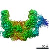
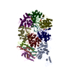
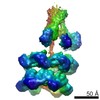
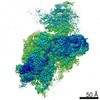
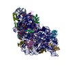
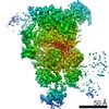
| Title | Cardiac thin filament decorated with C0C1 fragment of cardiac myosin binding protein C mode 1 | ||||||
 Components Components |
| ||||||
 Keywords Keywords | MOTOR PROTEIN / myosin binding protein C | ||||||
| Function / homology |  Function and homology information Function and homology informationbasal body patch / C zone / regulation of muscle filament sliding / striated muscle myosin thick filament / tight junction assembly / regulation of striated muscle contraction / cardiac myofibril / profilin binding / A band / protein localization to bicellular tight junction ...basal body patch / C zone / regulation of muscle filament sliding / striated muscle myosin thick filament / tight junction assembly / regulation of striated muscle contraction / cardiac myofibril / profilin binding / A band / protein localization to bicellular tight junction / regulation of transepithelial transport / morphogenesis of a polarized epithelium / structural constituent of postsynaptic actin cytoskeleton / Formation of annular gap junctions / Formation of the dystrophin-glycoprotein complex (DGC) / Gap junction degradation / Cell-extracellular matrix interactions / dense body / Striated Muscle Contraction / regulation of stress fiber assembly / Regulation of CDH1 Function / regulation of cardiac muscle cell contraction / Adherens junctions interactions / M band / Sensory processing of sound by outer hair cells of the cochlea / regulation of focal adhesion assembly / Sensory processing of sound by inner hair cells of the cochlea / Interaction between L1 and Ankyrins / structural constituent of muscle / sarcomere organization / apical junction complex / ventricular cardiac muscle tissue morphogenesis / positive regulation of wound healing / myosin heavy chain binding / myosin binding / maintenance of blood-brain barrier / filamentous actin / NuA4 histone acetyltransferase complex / Recycling pathway of L1 / ATPase activator activity / myofibril / EPH-ephrin mediated repulsion of cells / regulation of synaptic vesicle endocytosis / RHO GTPases Activate WASPs and WAVEs / RHOBTB2 GTPase cycle / RHO GTPases activate IQGAPs / heart morphogenesis / phagocytic vesicle / cardiac muscle contraction / titin binding / EPHB-mediated forward signaling / axonogenesis / calyx of Held / sarcomere / FCGR3A-mediated phagocytosis / actin filament / Translocation of SLC2A4 (GLUT4) to the plasma membrane / cell motility / RHO GTPases Activate Formins / Signaling by high-kinase activity BRAF mutants / MAP2K and MAPK activation / Regulation of actin dynamics for phagocytic cup formation / structural constituent of cytoskeleton / VEGFA-VEGFR2 Pathway / platelet aggregation / cellular response to type II interferon / Hydrolases; Acting on acid anhydrides; Acting on acid anhydrides to facilitate cellular and subcellular movement / Schaffer collateral - CA1 synapse / Signaling by RAF1 mutants / Signaling by moderate kinase activity BRAF mutants / Paradoxical activation of RAF signaling by kinase inactive BRAF / Signaling downstream of RAS mutants / cell-cell junction / Signaling by BRAF and RAF1 fusions / actin cytoskeleton / Clathrin-mediated endocytosis / actin binding / angiogenesis / blood microparticle / cytoskeleton / cell adhesion / positive regulation of cell migration / axon / focal adhesion / hydrolase activity / ubiquitin protein ligase binding / positive regulation of gene expression / synapse / protein kinase binding / : / extracellular exosome / ATP binding / membrane / metal ion binding / identical protein binding / nucleus / plasma membrane / cytoplasm / cytosol Similarity search - Function | ||||||
| Biological species |  Homo sapiens (human) Homo sapiens (human) | ||||||
| Method | ELECTRON MICROSCOPY / helical reconstruction / cryo EM / Resolution: 11 Å | ||||||
 Authors Authors | Galkin, V.E. / Schroeder, G.F. | ||||||
| Funding support |  United States, 1items United States, 1items
| ||||||
 Citation Citation | Journal: Structure / Year: 2018 Title: N-Terminal Domains of Cardiac Myosin Binding Protein C Cooperatively Activate the Thin Filament. Authors: Cristina Risi / Betty Belknap / Eva Forgacs-Lonart / Samantha P Harris / Gunnar F Schröder / Howard D White / Vitold E Galkin /  Abstract: Muscle contraction relies on interaction between myosin-based thick filaments and actin-based thin filaments. Myosin binding protein C (MyBP-C) is a key regulator of actomyosin interactions. Recent ...Muscle contraction relies on interaction between myosin-based thick filaments and actin-based thin filaments. Myosin binding protein C (MyBP-C) is a key regulator of actomyosin interactions. Recent studies established that the N'-terminal domains (NTDs) of MyBP-C can either activate or inhibit thin filaments, but the mechanism of their collective action is poorly understood. Cardiac MyBP-C (cMyBP-C) harbors an extra NTD, which is absent in skeletal isoforms of MyBP-C, and its role in regulation of cardiac contraction is unknown. Here we show that the first two domains of human cMyPB-C (i.e., C0 and C1) cooperate to activate the thin filament. We demonstrate that C1 interacts with tropomyosin via a positively charged loop and that this interaction, stabilized by the C0 domain, is required for thin filament activation by cMyBP-C. Our data reveal a mechanism by which cMyBP-C can modulate cardiac contraction and demonstrate a function of the C0 domain. | ||||||
| History |
|
- Structure visualization
| Movie |
Movie viewer |
|---|---|
| Structure viewer | Molecule: MolmilJmol/JSmol |
- Downloads & links
-Download
| PDBx/mmCIF format | 6cxi.cif.gz | 533.7 KB | Display | PDBx/mmCIF format |
|---|---|---|---|---|
| PDB format | pdb6cxi.ent.gz | 428.4 KB | Display | PDB format |
| PDBx/mmJSON format | 6cxi.json.gz | Tree view | PDBx/mmJSON format | |
| Others |  Other downloads Other downloads |
-Validation report
| Arichive directory | https://data.pdbj.org/pub/pdb/validation_reports/cx/6cxiftp://data.pdbj.org/pub/pdb/validation_reports/cx/6cxi | HTTPS FTP |
|---|
-Related structure data
| Related structure data | 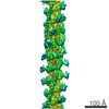 7780MC 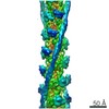 4346C 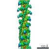 7781C 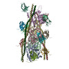 6cxjC 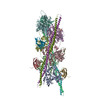 6g2tC M: map data used to model this data C: citing same article ( |
|---|---|
| Similar structure data |


-Links
 PDBj
PDBj


- Assembly
| Deposited unit | 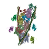
|
|---|---|
| 1 |
|
-Components
| #1: Protein | Mass: 41838.766 Da / Num. of mol.: 5 Source method: isolated from a genetically manipulated source Source: (gene. exp.) Homo sapiens (human) / Gene: ACTG1, ACTG / Production host: Homo sapiens (human) / References: UniProt: P63261#2: Protein | Mass: 12180.806 Da / Num. of mol.: 6 / Fragment: C1 Ig-domain (UNP residues 151-258) Source method: isolated from a genetically manipulated source Source: (gene. exp.) Homo sapiens (human) / Gene: MYBPC3 / Production host:  #3: Protein | Mass: 10706.060 Da / Num. of mol.: 5 / Fragment: C0 Ig-domain (UNP residues 1-101) Source method: isolated from a genetically manipulated source Source: (gene. exp.) Homo sapiens (human) / Gene: MYBPC3 / Production host: #4: Protein | Mass: 10826.337 Da / Num. of mol.: 4 Source method: isolated from a genetically manipulated source Source: (gene. exp.) Homo sapiens (human) / Production host: Homo sapiens (human) |
|---|
-Experimental details
-Experiment
| Experiment | Method: ELECTRON MICROSCOPY |
|---|---|
| EM experiment | Aggregation state: HELICAL ARRAY / 3D reconstruction method: helical reconstruction |
- Sample preparation
| Component |
| |||||||||||||||||||||||||||||||||||
|---|---|---|---|---|---|---|---|---|---|---|---|---|---|---|---|---|---|---|---|---|---|---|---|---|---|---|---|---|---|---|---|---|---|---|---|---|
| Molecular weight | Experimental value: NO | |||||||||||||||||||||||||||||||||||
| Source (natural) |
| |||||||||||||||||||||||||||||||||||
| Source (recombinant) |
| |||||||||||||||||||||||||||||||||||
| Buffer solution | pH: 7 | |||||||||||||||||||||||||||||||||||
| Specimen | Embedding applied: NO / Shadowing applied: NO / Staining applied: NO / Vitrification applied: YES | |||||||||||||||||||||||||||||||||||
| Specimen support | Grid material: COPPER / Grid mesh size: 300 divisions/in. | |||||||||||||||||||||||||||||||||||
| Vitrification | Instrument: FEI VITROBOT MARK II / Cryogen name: ETHANE / Humidity: 95 % / Chamber temperature: 294 K |
- Electron microscopy imaging
| Experimental equipment |  Model: Titan Krios / Image courtesy: FEI Company |
|---|---|
| Microscopy | Model: FEI TITAN KRIOS |
| Electron gun | Electron source:  FIELD EMISSION GUN / Accelerating voltage: 300 kV / Illumination mode: FLOOD BEAM FIELD EMISSION GUN / Accelerating voltage: 300 kV / Illumination mode: FLOOD BEAM |
| Electron lens | Mode: BRIGHT FIELD |
| Image recording | Electron dose: 20 e/Å2 / Detector mode: INTEGRATING / Film or detector model: FEI FALCON II (4k x 4k) |
- Processing
| EM software |
| ||||||||||||||||||||||||||||||||||||
|---|---|---|---|---|---|---|---|---|---|---|---|---|---|---|---|---|---|---|---|---|---|---|---|---|---|---|---|---|---|---|---|---|---|---|---|---|---|
| CTF correction | Type: PHASE FLIPPING AND AMPLITUDE CORRECTION | ||||||||||||||||||||||||||||||||||||
| Helical symmerty | Angular rotation/subunit: -166.6 ° / Axial rise/subunit: 27.5 Å / Axial symmetry: C1 | ||||||||||||||||||||||||||||||||||||
| 3D reconstruction | Resolution: 11 Å / Resolution method: FSC 0.143 CUT-OFF / Num. of particles: 6117 / Algorithm: BACK PROJECTION / Symmetry type: HELICAL | ||||||||||||||||||||||||||||||||||||
| Atomic model building | Protocol: FLEXIBLE FIT / Space: REAL / Target criteria: Correlation coefficient |
