[English] 日本語
 Yorodumi
- PDB-6cwa: CRYSTAL STRUCTURE PHGDH IN COMPLEX WITH NADH AND 3-PHOSPHOGLYCERA... -
+ Open data
- Basic information
| Entry | Database: PDB / ID: 6cwa | ||||||
|---|---|---|---|---|---|---|---|
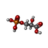
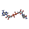
| Title | CRYSTAL STRUCTURE PHGDH IN COMPLEX WITH NADH AND 3-PHOSPHOGLYCERATE AT 1.77 A RESOLUTION | ||||||
 Components Components | D-3-phosphoglycerate dehydrogenase | ||||||
 Keywords Keywords | OXIDOREDUCTASE / PHGDH | ||||||
| Function / homology |  Function and homology information Function and homology information2-oxoglutarate reductase / threonine metabolic process / glial cell development / taurine metabolic process / phosphoglycerate dehydrogenase / phosphoglycerate dehydrogenase activity / gamma-aminobutyric acid metabolic process / Serine metabolism / glycine metabolic process / L-serine biosynthetic process ...2-oxoglutarate reductase / threonine metabolic process / glial cell development / taurine metabolic process / phosphoglycerate dehydrogenase / phosphoglycerate dehydrogenase activity / gamma-aminobutyric acid metabolic process / Serine metabolism / glycine metabolic process / L-serine biosynthetic process / malate dehydrogenase / L-malate dehydrogenase (NAD+) activity / glutamine metabolic process / G1 to G0 transition / neural tube development / spinal cord development / brain development / NAD binding / neuron projection development / regulation of gene expression / electron transfer activity / extracellular exosome / cytosol Similarity search - Function | ||||||
| Biological species |  Homo sapiens (human) Homo sapiens (human) | ||||||
| Method |  X-RAY DIFFRACTION / X-RAY DIFFRACTION /  SYNCHROTRON / SYNCHROTRON /  MOLECULAR REPLACEMENT / Resolution: 1.77 Å MOLECULAR REPLACEMENT / Resolution: 1.77 Å | ||||||
 Authors Authors | Davies, D.R. / Edwards, T.E. | ||||||
 Citation Citation |  Journal: J.Med.Chem. / Year: 2019 Journal: J.Med.Chem. / Year: 2019Title: Intracellular Trapping of the Selective Phosphoglycerate Dehydrogenase (PHGDH) InhibitorBI-4924Disrupts Serine Biosynthesis. Authors: Weinstabl, H. / Treu, M. / Rinnenthal, J. / Zahn, S.K. / Ettmayer, P. / Bader, G. / Dahmann, G. / Kessler, D. / Rumpel, K. / Mischerikow, N. / Savarese, F. / Gerstberger, T. / Mayer, M. / ...Authors: Weinstabl, H. / Treu, M. / Rinnenthal, J. / Zahn, S.K. / Ettmayer, P. / Bader, G. / Dahmann, G. / Kessler, D. / Rumpel, K. / Mischerikow, N. / Savarese, F. / Gerstberger, T. / Mayer, M. / Zoephel, A. / Schnitzer, R. / Sommergruber, W. / Martinelli, P. / Arnhof, H. / Peric-Simov, B. / Hofbauer, K.S. / Garavel, G. / Scherbantin, Y. / Mitzner, S. / Fett, T.N. / Scholz, G. / Bruchhaus, J. / Burkard, M. / Kousek, R. / Ciftci, T. / Sharps, B. / Schrenk, A. / Harrer, C. / Haering, D. / Wolkerstorfer, B. / Zhang, X. / Lv, X. / Du, A. / Li, D. / Li, Y. / Quant, J. / Pearson, M. / McConnell, D.B. | ||||||
| History |
|
- Structure visualization
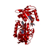
| Structure viewer | Molecule:  Molmil Molmil Jmol/JSmol Jmol/JSmol |
|---|
- Downloads & links
- Download
| PDBx/mmCIF format |  6cwa.cif.gz 6cwa.cif.gz | 231.9 KB | Display |  PDBx/mmCIF format PDBx/mmCIF format |
|---|---|---|---|---|
| PDB format |  pdb6cwa.ent.gz pdb6cwa.ent.gz | 183.1 KB | Display |  PDB format PDB format |
| PDBx/mmJSON format |  6cwa.json.gz 6cwa.json.gz | Tree view |  PDBx/mmJSON format PDBx/mmJSON format | |
| Others |  Other downloads Other downloads |
-Validation report
| Summary document |  6cwa_validation.pdf.gz 6cwa_validation.pdf.gz | 1006.6 KB | Display |  wwPDB validaton report wwPDB validaton report |
|---|---|---|---|---|
| Full document |  6cwa_full_validation.pdf.gz 6cwa_full_validation.pdf.gz | 1021.4 KB | Display | |
| Data in XML |  6cwa_validation.xml.gz 6cwa_validation.xml.gz | 27.2 KB | Display | |
| Data in CIF |  6cwa_validation.cif.gz 6cwa_validation.cif.gz | 38 KB | Display | |
| Arichive directory |  https://data.pdbj.org/pub/pdb/validation_reports/cw/6cwa https://data.pdbj.org/pub/pdb/validation_reports/cw/6cwa ftp://data.pdbj.org/pub/pdb/validation_reports/cw/6cwa ftp://data.pdbj.org/pub/pdb/validation_reports/cw/6cwa | HTTPS FTP |
-Related structure data
| Related structure data | 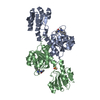 6rihC 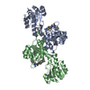 6rj2C 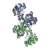 6rj3C 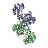 6rj5C 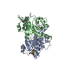 6rj6C 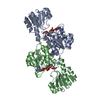 2g76S S: Starting model for refinement C: citing same article ( |
|---|---|
| Similar structure data |
- Links
Links
- Assembly
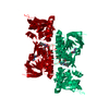
| Deposited unit | 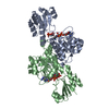
| ||||||||
|---|---|---|---|---|---|---|---|---|---|
| 1 |
| ||||||||
| Unit cell |
|
- Components
| #1: Protein | Mass: 35055.266 Da / Num. of mol.: 2 / Fragment: PHGDH Source method: isolated from a genetically manipulated source Source: (gene. exp.)  Homo sapiens (human) / Gene: PHGDH, PGDH3 / Production host: Homo sapiens (human) / Gene: PHGDH, PGDH3 / Production host:  References: UniProt: O43175, phosphoglycerate dehydrogenase, 2-oxoglutarate reductase, malate dehydrogenase #2: Chemical | #3: Chemical | #4: Water | ChemComp-HOH / | |
|---|
-Experimental details
-Experiment
| Experiment | Method:  X-RAY DIFFRACTION / Number of used crystals: 1 X-RAY DIFFRACTION / Number of used crystals: 1 |
|---|
- Sample preparation
| Crystal | Density Matthews: 2.29 Å3/Da / Density % sol: 46.22 % |
|---|---|
| Crystal grow | Temperature: 289 K / Method: vapor diffusion, sitting drop / pH: 6.5 Details: 100 MM IMIDAZOLE PH 6.5, 20% PEG 3350, 100 MM MAGNESIUM CHLORIDE, 2M NACL, 1MM NADH; SOAK OVERNIGHT IN CRYSTALLIZATION SOLUTION PLUS 5 MM 3- PHOSPHOGLYCERATE; 20% EG CRYO, VAPOR DIFFUSION, TEMPERATURE 289K PH range: 6.5 |
-Data collection
| Diffraction | Mean temperature: 100 K |
|---|---|
| Diffraction source | Source:  SYNCHROTRON / Site: SYNCHROTRON / Site:  APS APS  / Beamline: 21-ID-F / Wavelength: 0.97872 Å / Beamline: 21-ID-F / Wavelength: 0.97872 Å |
| Detector | Type: MARMOSAIC 225 mm CCD / Detector: CCD / Date: Jun 12, 2014 |
| Radiation | Protocol: SINGLE WAVELENGTH / Monochromatic (M) / Laue (L): M / Scattering type: x-ray |
| Radiation wavelength | Wavelength: 0.97872 Å / Relative weight: 1 |
| Reflection | Resolution: 1.77→28.36 Å / Num. obs: 58407 / % possible obs: 96.5 % / Observed criterion σ(I): -3 / Redundancy: 2.65 % / Biso Wilson estimate: 28.9 Å2 / Rmerge(I) obs: 0.05 / Net I/σ(I): 15.19 |
| Reflection shell | Resolution: 1.77→1.82 Å / Rmerge(I) obs: 0.5 / Mean I/σ(I) obs: 2.8 / % possible all: 95.1 |
- Processing
| Software |
| ||||||||||||||||||||||||||||||||||||||||||||||||||||||||||||||||||||||||||||||||||||||||||||||||||||||||||||||||||||||||||||||||||||||||||||||||||||||
|---|---|---|---|---|---|---|---|---|---|---|---|---|---|---|---|---|---|---|---|---|---|---|---|---|---|---|---|---|---|---|---|---|---|---|---|---|---|---|---|---|---|---|---|---|---|---|---|---|---|---|---|---|---|---|---|---|---|---|---|---|---|---|---|---|---|---|---|---|---|---|---|---|---|---|---|---|---|---|---|---|---|---|---|---|---|---|---|---|---|---|---|---|---|---|---|---|---|---|---|---|---|---|---|---|---|---|---|---|---|---|---|---|---|---|---|---|---|---|---|---|---|---|---|---|---|---|---|---|---|---|---|---|---|---|---|---|---|---|---|---|---|---|---|---|---|---|---|---|---|---|---|
| Refinement | Method to determine structure:  MOLECULAR REPLACEMENT MOLECULAR REPLACEMENTStarting model: 2G76 Resolution: 1.77→28.36 Å / Cross valid method: FREE R-VALUE
| ||||||||||||||||||||||||||||||||||||||||||||||||||||||||||||||||||||||||||||||||||||||||||||||||||||||||||||||||||||||||||||||||||||||||||||||||||||||
| Solvent computation | Shrinkage radii: 0.9 Å | ||||||||||||||||||||||||||||||||||||||||||||||||||||||||||||||||||||||||||||||||||||||||||||||||||||||||||||||||||||||||||||||||||||||||||||||||||||||
| Displacement parameters | Biso mean: 36.59 Å2 | ||||||||||||||||||||||||||||||||||||||||||||||||||||||||||||||||||||||||||||||||||||||||||||||||||||||||||||||||||||||||||||||||||||||||||||||||||||||
| Refinement step | Cycle: LAST / Resolution: 1.77→28.36 Å
| ||||||||||||||||||||||||||||||||||||||||||||||||||||||||||||||||||||||||||||||||||||||||||||||||||||||||||||||||||||||||||||||||||||||||||||||||||||||
| Refinement TLS params. | Method: refined / Refine-ID: X-RAY DIFFRACTION
|
 Movie
Movie Controller



 PDBj
PDBj


