 Movie
Movie Controller

[English] 日本語
 Yorodumi
- PDB-6cbt: DnaG Primase C-terminal domain complex with SSB C-terminal peptide -
+ Open data

- Basic information
| Entry | Database: PDB / ID: 6cbt | ||||||
|---|---|---|---|---|---|---|---|
| Title | DnaG Primase C-terminal domain complex with SSB C-terminal peptide | ||||||
 Components Components | DNA primase, single-stranded DNA-binding protein chimera | ||||||
 Keywords Keywords | REPLICATION / Primase / DnaG / Single-strand DNA-Binding Protein / SSB / Complex | ||||||
| Function / homology |  Function and homology information Function and homology informationDnaB-DnaG complex / DNA primase DnaG / primosome complex / DNA replication, synthesis of primer / replisome / replication fork processing / DNA-directed RNA polymerase complex / DNA-directed RNA polymerase activity / single-stranded DNA binding / DNA recombination ...DnaB-DnaG complex / DNA primase DnaG / primosome complex / DNA replication, synthesis of primer / replisome / replication fork processing / DNA-directed RNA polymerase complex / DNA-directed RNA polymerase activity / single-stranded DNA binding / DNA recombination / DNA replication / DNA repair / DNA binding / zinc ion binding / cytoplasm Similarity search - Function | ||||||
| Biological species |  | ||||||
| Method |  X-RAY DIFFRACTION / SYNCHROTRON / MOLECULAR REPLACEMENT / Resolution: 2.1 Å X-RAY DIFFRACTION / SYNCHROTRON / MOLECULAR REPLACEMENT / Resolution: 2.1 Å | ||||||
 Authors Authors | Oakley, A.J. / Lo, A.T.Y. | ||||||
| Funding support |  Australia, 1items Australia, 1items
| ||||||
 Citation Citation | Journal: To be Published Title: DnaG Primase C-terminal domain complex with SSB C-terminal peptide Authors: Oakley, A.J. / Lo, A.T.Y. | ||||||
| History |
|
- Structure visualization
| Structure viewer | Molecule: MolmilJmol/JSmol |
|---|
- Downloads & links
-Download
| PDBx/mmCIF format | 6cbt.cif.gz | 136.2 KB | Display | PDBx/mmCIF format |
|---|---|---|---|---|
| PDB format | pdb6cbt.ent.gz | 104.3 KB | Display | PDB format |
| PDBx/mmJSON format | 6cbt.json.gz | Tree view | PDBx/mmJSON format | |
| Others |  Other downloads Other downloads |
-Validation report
| Arichive directory | https://data.pdbj.org/pub/pdb/validation_reports/cb/6cbtftp://data.pdbj.org/pub/pdb/validation_reports/cb/6cbt | HTTPS FTP |
|---|
-Related structure data
| Related structure data |  6cbrC  6cbsC  1t3wS S: Starting model for refinement C: citing same article ( |
|---|---|
| Similar structure data |


-Links
 PDBj
PDBj


- Assembly
| Deposited unit | 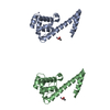
| |||||||||
|---|---|---|---|---|---|---|---|---|---|---|
| 1 | 
| |||||||||
| 2 | 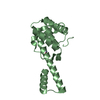
| |||||||||
| Unit cell |
| |||||||||
| Components on special symmetry positions |
|

-Components
| #1: Protein | Mass: 18691.049 Da / Num. of mol.: 2 Fragment: DnaG C-terminal domain (UNP residues 434-581), linker peptide, SSB C-terminal peptide (UNP residues 130-139) Source method: isolated from a genetically manipulated source Source: (gene. exp.) Strain: K12 Gene: dnaG, dnaP, parB, b3066, JW3038, ssb_1, BUE81_14300, ERS085374_00666 Plasmid: pAL1402 / Production host: #2: Chemical |   Mass: 65.409 Da / Num. of mol.: 3 / Source method: obtained synthetically / Formula: Zn Mass: 65.409 Da / Num. of mol.: 3 / Source method: obtained synthetically / Formula: Zn#3: Chemical |   Mass: 60.052 Da / Num. of mol.: 2 / Source method: obtained synthetically / Formula: C2H4O2 Mass: 60.052 Da / Num. of mol.: 2 / Source method: obtained synthetically / Formula: C2H4O2#4: Water | ChemComp-HOH / |  Mass: 18.015 Da / Num. of mol.: 310 / Source method: isolated from a natural source / Formula: H2O Mass: 18.015 Da / Num. of mol.: 310 / Source method: isolated from a natural source / Formula: H2O |
|---|
-Experimental details
-Experiment
| Experiment | Method: X-RAY DIFFRACTION / Number of used crystals: 1 |
|---|
- Sample preparation
| Crystal | Density Matthews: 2.49 Å3/Da / Density % sol: 50.62 % |
|---|---|
| Crystal grow | Temperature: 294 K / Method: vapor diffusion, hanging drop / pH: 4.6 Details: 9.5% w/v PEG3000, 10 mM zinc acetate, 100 mM sodium acetate, pH 4.6 |
-Data collection
| Diffraction | Mean temperature: 100 K |
|---|---|
| Diffraction source | Source: SYNCHROTRON / Site: Australian Synchrotron / Beamline: MX2 / Wavelength: 0.953715 Å |
| Detector | Type: ADSC QUANTUM 315r / Detector: CCD / Date: Aug 29, 2009 |
| Radiation | Protocol: SINGLE WAVELENGTH / Monochromatic (M) / Laue (L): M / Scattering type: x-ray |
| Radiation wavelength | Wavelength: 0.953715 Å / Relative weight: 1 |
| Reflection | Resolution: 2.1→70 Å / Num. obs: 21786 / % possible obs: 96.8 % / Redundancy: 6.3 % / Biso Wilson estimate: 22.702 Å2 / Rmerge(I) obs: 0.133 / Rpim(I) all: 0.057 / Rrim(I) all: 0.146 / Net I/σ(I): 9 |
| Reflection shell | Resolution: 2.1→2.21 Å / Redundancy: 6.4 % / Rmerge(I) obs: 0.56 / Mean I/σ(I) obs: 3.1 / Num. unique obs: 3075 / Rpim(I) all: 0.23 / Rrim(I) all: 0.61 / % possible all: 95.7 |
- Processing
| Software |
| |||||||||||||||||||||||||||||||||||||||||||||||||||||||||||||||||||||||||||
|---|---|---|---|---|---|---|---|---|---|---|---|---|---|---|---|---|---|---|---|---|---|---|---|---|---|---|---|---|---|---|---|---|---|---|---|---|---|---|---|---|---|---|---|---|---|---|---|---|---|---|---|---|---|---|---|---|---|---|---|---|---|---|---|---|---|---|---|---|---|---|---|---|---|---|---|---|
| Refinement | Method to determine structure: MOLECULAR REPLACEMENT Starting model: PDB entry 1T3W Resolution: 2.1→45.742 Å / SU ML: 0.23 / Cross valid method: THROUGHOUT / σ(F): 1.35 / Phase error: 23.2 / Stereochemistry target values: ML
| |||||||||||||||||||||||||||||||||||||||||||||||||||||||||||||||||||||||||||
| Solvent computation | Shrinkage radii: 0.9 Å / VDW probe radii: 1.11 Å / Solvent model: FLAT BULK SOLVENT MODEL | |||||||||||||||||||||||||||||||||||||||||||||||||||||||||||||||||||||||||||
| Refinement step | Cycle: LAST / Resolution: 2.1→45.742 Å
| |||||||||||||||||||||||||||||||||||||||||||||||||||||||||||||||||||||||||||
| Refine LS restraints |
| |||||||||||||||||||||||||||||||||||||||||||||||||||||||||||||||||||||||||||
| LS refinement shell |
| |||||||||||||||||||||||||||||||||||||||||||||||||||||||||||||||||||||||||||
| Refinement TLS params. | Method: refined / Refine-ID: X-RAY DIFFRACTION
| |||||||||||||||||||||||||||||||||||||||||||||||||||||||||||||||||||||||||||
| Refinement TLS group |
|
